Zinc »
PDB 2rgv-2rv4 »
2rkn »
Zinc in PDB 2rkn: X-Ray Structure of the Self-Defense and Signaling Protein DIR1 From Arabidopsis Taliana
Protein crystallography data
The structure of X-Ray Structure of the Self-Defense and Signaling Protein DIR1 From Arabidopsis Taliana, PDB code: 2rkn
was solved by
M.B.Lascombe,
T.Prange,
N.Buhot,
D.Marion,
B.Bakan,
C.Lamb,
with X-Ray Crystallography technique. A brief refinement statistics is given in the table below:
| Resolution Low / High (Å) | 9.92 / 1.60 |
| Space group | P 21 21 21 |
| Cell size a, b, c (Å), α, β, γ (°) | 28.655, 48.217, 54.406, 90.00, 90.00, 90.00 |
| R / Rfree (%) | 18.9 / 25.2 |
Zinc Binding Sites:
The binding sites of Zinc atom in the X-Ray Structure of the Self-Defense and Signaling Protein DIR1 From Arabidopsis Taliana
(pdb code 2rkn). This binding sites where shown within
5.0 Angstroms radius around Zinc atom.
In total 3 binding sites of Zinc where determined in the X-Ray Structure of the Self-Defense and Signaling Protein DIR1 From Arabidopsis Taliana, PDB code: 2rkn:
Jump to Zinc binding site number: 1; 2; 3;
In total 3 binding sites of Zinc where determined in the X-Ray Structure of the Self-Defense and Signaling Protein DIR1 From Arabidopsis Taliana, PDB code: 2rkn:
Jump to Zinc binding site number: 1; 2; 3;
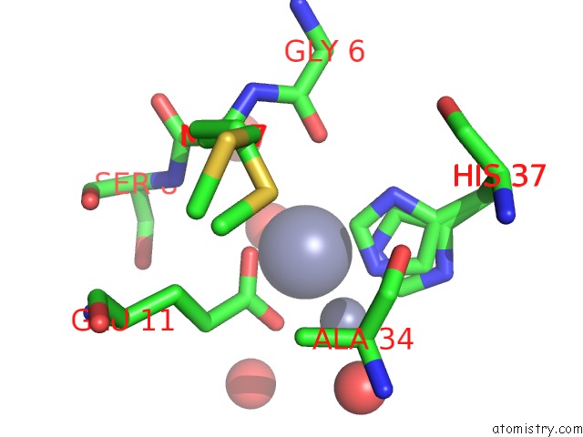
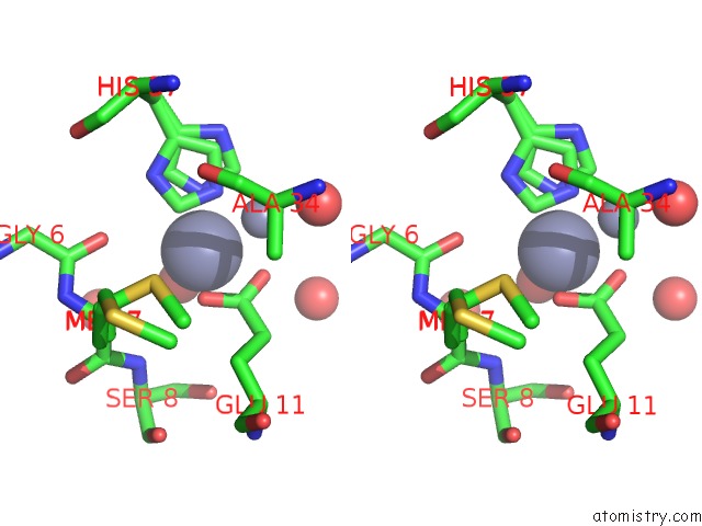
Zinc binding site 1 out of 3 in 2rkn
Go back to
Zinc binding site 1 out
of 3 in the X-Ray Structure of the Self-Defense and Signaling Protein DIR1 From Arabidopsis Taliana
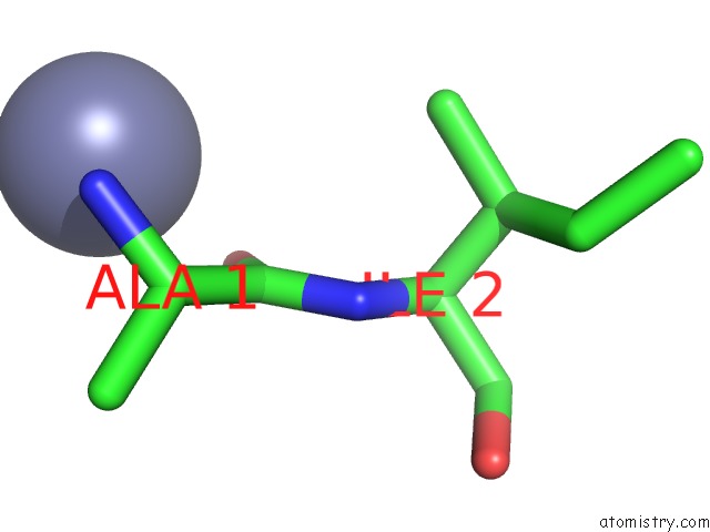
Mono view
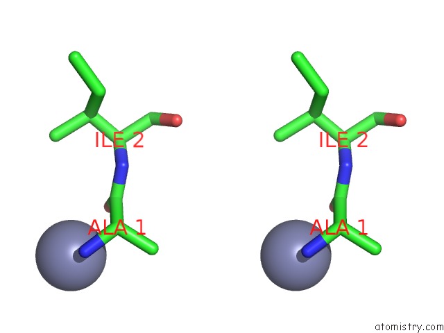
Stereo pair view
Mono view
Stereo pair view
A full contact list of Zinc with other atoms in the Zn binding
site number 1 of X-Ray Structure of the Self-Defense and Signaling Protein DIR1 From Arabidopsis Taliana within 5.0Å range:
|
Zinc binding site 2 out of 3 in 2rkn
Go back to
Zinc binding site 2 out
of 3 in the X-Ray Structure of the Self-Defense and Signaling Protein DIR1 From Arabidopsis Taliana
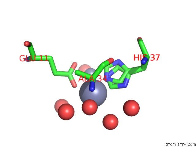
Mono view
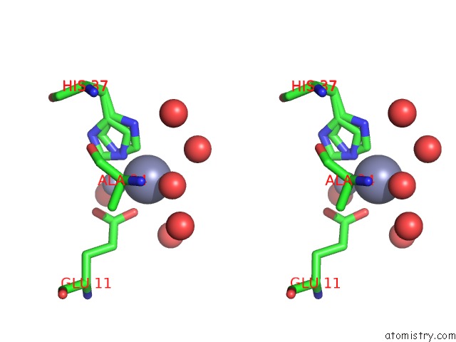
Stereo pair view
Mono view
Stereo pair view
A full contact list of Zinc with other atoms in the Zn binding
site number 2 of X-Ray Structure of the Self-Defense and Signaling Protein DIR1 From Arabidopsis Taliana within 5.0Å range:
|
Zinc binding site 3 out of 3 in 2rkn
Go back to
Zinc binding site 3 out
of 3 in the X-Ray Structure of the Self-Defense and Signaling Protein DIR1 From Arabidopsis Taliana
Mono view
Stereo pair view
Mono view
Stereo pair view
A full contact list of Zinc with other atoms in the Zn binding
site number 3 of X-Ray Structure of the Self-Defense and Signaling Protein DIR1 From Arabidopsis Taliana within 5.0Å range:
|
Reference:
M.B.Lascombe,
B.Bakan,
N.Buhot,
D.Marion,
J.P.Blein,
V.Larue,
C.Lamb,
T.Prange.
The Structure of "Defective in Induced Resistance" Protein of Arabidopsis Thaliana, DIR1, Reveals A New Type of Lipid Transfer Protein. Protein Sci. V. 17 1522 2008.
ISSN: ISSN 0961-8368
PubMed: 18552128
DOI: 10.1110/PS.035972.108
Page generated: Thu Oct 17 03:50:35 2024
ISSN: ISSN 0961-8368
PubMed: 18552128
DOI: 10.1110/PS.035972.108
Last articles
Zn in 9MJ5Zn in 9HNW
Zn in 9G0L
Zn in 9FNE
Zn in 9DZN
Zn in 9E0I
Zn in 9D32
Zn in 9DAK
Zn in 8ZXC
Zn in 8ZUF