Zinc »
PDB 2qmy-2qzr »
2qyv »
Zinc in PDB 2qyv: Crystal Structure of Putative Xaa-His Dipeptidase (YP_718209.1) From Haemophilus Somnus 129PT at 2.11 A Resolution
Enzymatic activity of Crystal Structure of Putative Xaa-His Dipeptidase (YP_718209.1) From Haemophilus Somnus 129PT at 2.11 A Resolution
All present enzymatic activity of Crystal Structure of Putative Xaa-His Dipeptidase (YP_718209.1) From Haemophilus Somnus 129PT at 2.11 A Resolution:
3.4.13.20;
3.4.13.20;
Protein crystallography data
The structure of Crystal Structure of Putative Xaa-His Dipeptidase (YP_718209.1) From Haemophilus Somnus 129PT at 2.11 A Resolution, PDB code: 2qyv
was solved by
Joint Center For Structural Genomics (Jcsg),
with X-Ray Crystallography technique. A brief refinement statistics is given in the table below:
| Resolution Low / High (Å) | 47.84 / 2.11 |
| Space group | P 21 21 2 |
| Cell size a, b, c (Å), α, β, γ (°) | 173.922, 84.293, 123.204, 90.00, 90.00, 90.00 |
| R / Rfree (%) | 22 / 24.4 |
Zinc Binding Sites:
The binding sites of Zinc atom in the Crystal Structure of Putative Xaa-His Dipeptidase (YP_718209.1) From Haemophilus Somnus 129PT at 2.11 A Resolution
(pdb code 2qyv). This binding sites where shown within
5.0 Angstroms radius around Zinc atom.
In total 4 binding sites of Zinc where determined in the Crystal Structure of Putative Xaa-His Dipeptidase (YP_718209.1) From Haemophilus Somnus 129PT at 2.11 A Resolution, PDB code: 2qyv:
Jump to Zinc binding site number: 1; 2; 3; 4;
In total 4 binding sites of Zinc where determined in the Crystal Structure of Putative Xaa-His Dipeptidase (YP_718209.1) From Haemophilus Somnus 129PT at 2.11 A Resolution, PDB code: 2qyv:
Jump to Zinc binding site number: 1; 2; 3; 4;
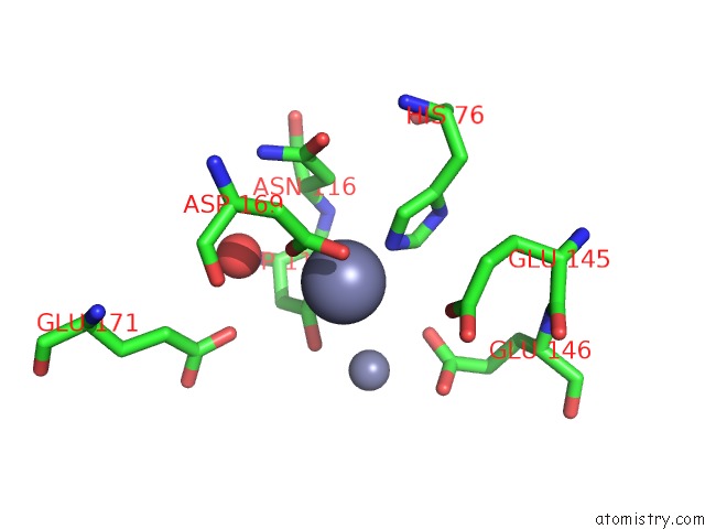
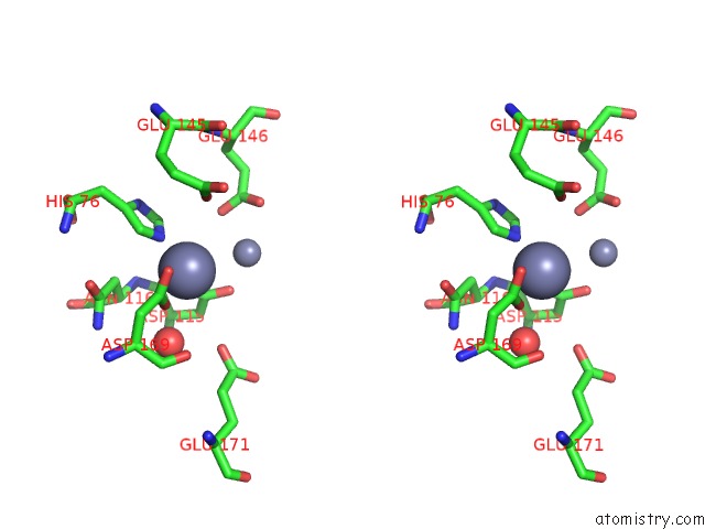
Zinc binding site 1 out of 4 in 2qyv
Go back to
Zinc binding site 1 out
of 4 in the Crystal Structure of Putative Xaa-His Dipeptidase (YP_718209.1) From Haemophilus Somnus 129PT at 2.11 A Resolution
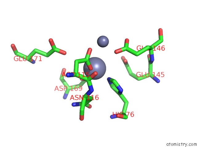
Mono view
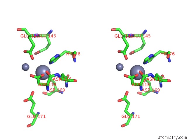
Stereo pair view
Mono view
Stereo pair view
A full contact list of Zinc with other atoms in the Zn binding
site number 1 of Crystal Structure of Putative Xaa-His Dipeptidase (YP_718209.1) From Haemophilus Somnus 129PT at 2.11 A Resolution within 5.0Å range:
|
Zinc binding site 2 out of 4 in 2qyv
Go back to
Zinc binding site 2 out
of 4 in the Crystal Structure of Putative Xaa-His Dipeptidase (YP_718209.1) From Haemophilus Somnus 129PT at 2.11 A Resolution
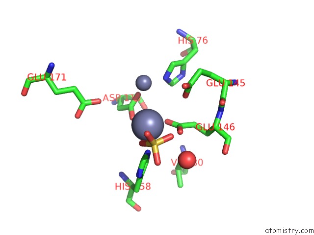
Mono view
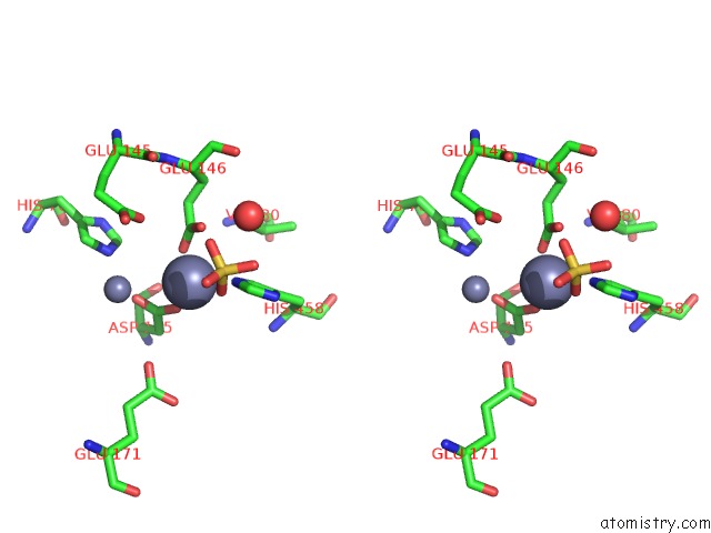
Stereo pair view
Mono view
Stereo pair view
A full contact list of Zinc with other atoms in the Zn binding
site number 2 of Crystal Structure of Putative Xaa-His Dipeptidase (YP_718209.1) From Haemophilus Somnus 129PT at 2.11 A Resolution within 5.0Å range:
|
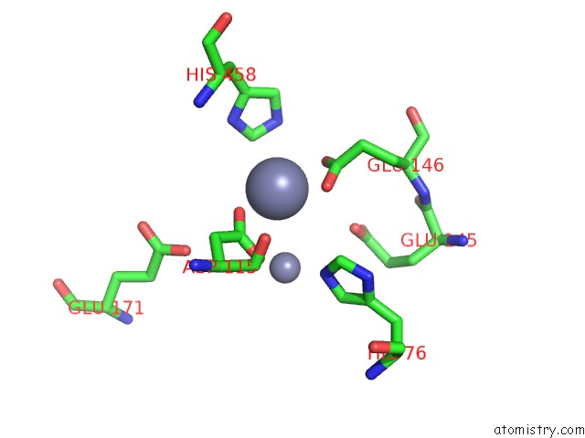
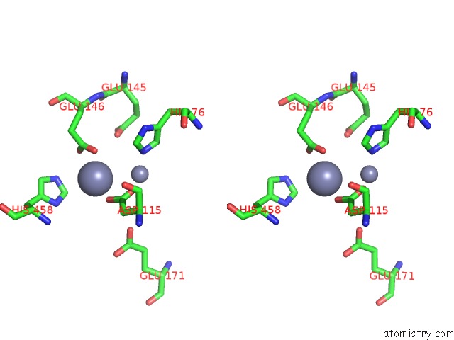
Zinc binding site 3 out of 4 in 2qyv
Go back to
Zinc binding site 3 out
of 4 in the Crystal Structure of Putative Xaa-His Dipeptidase (YP_718209.1) From Haemophilus Somnus 129PT at 2.11 A Resolution
Mono view
Stereo pair view
Mono view
Stereo pair view
A full contact list of Zinc with other atoms in the Zn binding
site number 3 of Crystal Structure of Putative Xaa-His Dipeptidase (YP_718209.1) From Haemophilus Somnus 129PT at 2.11 A Resolution within 5.0Å range:
|
Zinc binding site 4 out of 4 in 2qyv
Go back to
Zinc binding site 4 out
of 4 in the Crystal Structure of Putative Xaa-His Dipeptidase (YP_718209.1) From Haemophilus Somnus 129PT at 2.11 A Resolution
Mono view
Stereo pair view
Mono view
Stereo pair view
A full contact list of Zinc with other atoms in the Zn binding
site number 4 of Crystal Structure of Putative Xaa-His Dipeptidase (YP_718209.1) From Haemophilus Somnus 129PT at 2.11 A Resolution within 5.0Å range:
|
Reference:
Joint Center For Structural Genomics (Jcsg),
Joint Center For Structural Genomics (Jcsg).
N/A N/A.
Page generated: Wed Aug 20 05:31:35 2025
Last articles
Zn in 3HXEZn in 3HXD
Zn in 3HXC
Zn in 3HWP
Zn in 3HW7
Zn in 3HXB
Zn in 3HUV
Zn in 3HUD
Zn in 3HSV
Zn in 3HTR