Zinc »
PDB 2q42-2qmu »
2qa1 »
Zinc in PDB 2qa1: Crystal Structure of Pgae, An Aromatic Hydroxylase Involved in Angucycline Biosynthesis
Protein crystallography data
The structure of Crystal Structure of Pgae, An Aromatic Hydroxylase Involved in Angucycline Biosynthesis, PDB code: 2qa1
was solved by
H.Koskiniemi,
D.Dobritzsch,
M.Metsa-Ketela,
P.Kallio,
J.Niemi,
G.Schneider,
with X-Ray Crystallography technique. A brief refinement statistics is given in the table below:
| Resolution Low / High (Å) | 36.96 / 1.80 |
| Space group | F 2 2 2 |
| Cell size a, b, c (Å), α, β, γ (°) | 66.229, 171.535, 212.186, 90.00, 90.00, 90.00 |
| R / Rfree (%) | 19.5 / 22.4 |
Zinc Binding Sites:
The binding sites of Zinc atom in the Crystal Structure of Pgae, An Aromatic Hydroxylase Involved in Angucycline Biosynthesis
(pdb code 2qa1). This binding sites where shown within
5.0 Angstroms radius around Zinc atom.
In total only one binding site of Zinc was determined in the Crystal Structure of Pgae, An Aromatic Hydroxylase Involved in Angucycline Biosynthesis, PDB code: 2qa1:
In total only one binding site of Zinc was determined in the Crystal Structure of Pgae, An Aromatic Hydroxylase Involved in Angucycline Biosynthesis, PDB code: 2qa1:
Zinc binding site 1 out of 1 in 2qa1
Go back to
Zinc binding site 1 out
of 1 in the Crystal Structure of Pgae, An Aromatic Hydroxylase Involved in Angucycline Biosynthesis
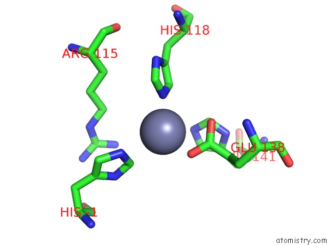
Mono view
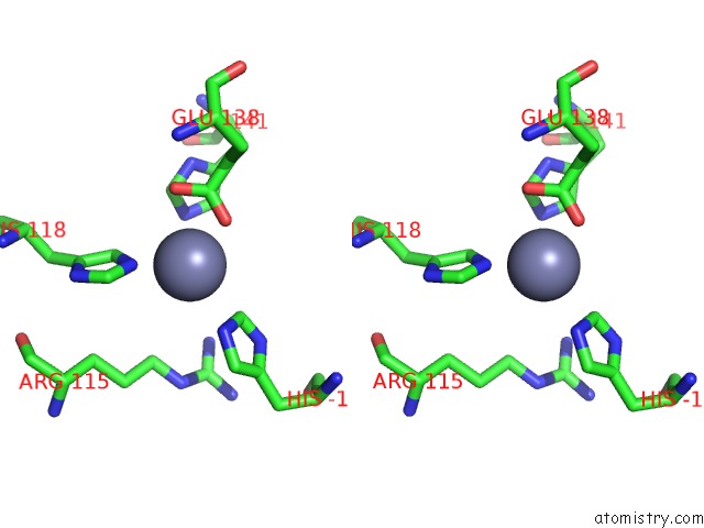
Stereo pair view
Mono view
Stereo pair view
A full contact list of Zinc with other atoms in the Zn binding
site number 1 of Crystal Structure of Pgae, An Aromatic Hydroxylase Involved in Angucycline Biosynthesis within 5.0Å range:
|
Reference:
H.Koskiniemi,
M.Metsa-Ketela,
D.Dobritzsch,
P.Kallio,
H.Korhonen,
P.Mantsala,
G.Schneider,
J.Niemi.
Crystal Structures of Two Aromatic Hydroxylases Involved in the Early Tailoring Steps of Angucycline Biosynthesis J.Mol.Biol. V. 372 633 2007.
ISSN: ISSN 0022-2836
PubMed: 17669423
DOI: 10.1016/J.JMB.2007.06.087
Page generated: Wed Aug 20 05:21:20 2025
ISSN: ISSN 0022-2836
PubMed: 17669423
DOI: 10.1016/J.JMB.2007.06.087
Last articles
Zn in 3GZ0Zn in 3GV4
Zn in 3GWT
Zn in 3GUC
Zn in 3GUG
Zn in 3GTV
Zn in 3GTT
Zn in 3GTQ
Zn in 3GTM
Zn in 3GTP