Zinc »
PDB 2pou-2q2l »
2pvv »
Zinc in PDB 2pvv: Structure of Human Glutamate Carboxypeptidase II (Gcpii) in Complex with L-Serine-O-Sulfate
Enzymatic activity of Structure of Human Glutamate Carboxypeptidase II (Gcpii) in Complex with L-Serine-O-Sulfate
All present enzymatic activity of Structure of Human Glutamate Carboxypeptidase II (Gcpii) in Complex with L-Serine-O-Sulfate:
3.4.17.21;
3.4.17.21;
Protein crystallography data
The structure of Structure of Human Glutamate Carboxypeptidase II (Gcpii) in Complex with L-Serine-O-Sulfate, PDB code: 2pvv
was solved by
C.Barinka,
J.Lubkowski,
with X-Ray Crystallography technique. A brief refinement statistics is given in the table below:
| Resolution Low / High (Å) | 30.00 / 2.11 |
| Space group | I 2 2 2 |
| Cell size a, b, c (Å), α, β, γ (°) | 101.617, 130.220, 158.975, 90.00, 90.00, 90.00 |
| R / Rfree (%) | 17.4 / 21.2 |
Other elements in 2pvv:
The structure of Structure of Human Glutamate Carboxypeptidase II (Gcpii) in Complex with L-Serine-O-Sulfate also contains other interesting chemical elements:
| Chlorine | (Cl) | 1 atom |
| Calcium | (Ca) | 1 atom |
Zinc Binding Sites:
The binding sites of Zinc atom in the Structure of Human Glutamate Carboxypeptidase II (Gcpii) in Complex with L-Serine-O-Sulfate
(pdb code 2pvv). This binding sites where shown within
5.0 Angstroms radius around Zinc atom.
In total 2 binding sites of Zinc where determined in the Structure of Human Glutamate Carboxypeptidase II (Gcpii) in Complex with L-Serine-O-Sulfate, PDB code: 2pvv:
Jump to Zinc binding site number: 1; 2;
In total 2 binding sites of Zinc where determined in the Structure of Human Glutamate Carboxypeptidase II (Gcpii) in Complex with L-Serine-O-Sulfate, PDB code: 2pvv:
Jump to Zinc binding site number: 1; 2;
Zinc binding site 1 out of 2 in 2pvv
Go back to
Zinc binding site 1 out
of 2 in the Structure of Human Glutamate Carboxypeptidase II (Gcpii) in Complex with L-Serine-O-Sulfate
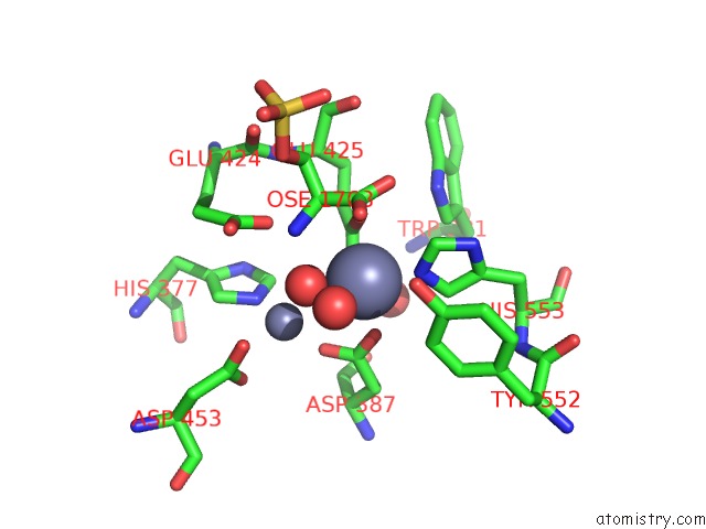
Mono view
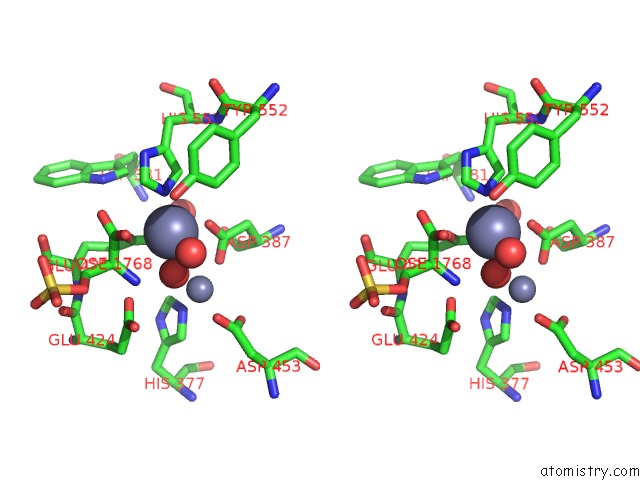
Stereo pair view
Mono view
Stereo pair view
A full contact list of Zinc with other atoms in the Zn binding
site number 1 of Structure of Human Glutamate Carboxypeptidase II (Gcpii) in Complex with L-Serine-O-Sulfate within 5.0Å range:
|
Zinc binding site 2 out of 2 in 2pvv
Go back to
Zinc binding site 2 out
of 2 in the Structure of Human Glutamate Carboxypeptidase II (Gcpii) in Complex with L-Serine-O-Sulfate
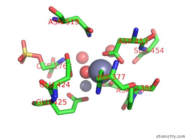
Mono view
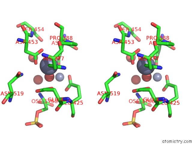
Stereo pair view
Mono view
Stereo pair view
A full contact list of Zinc with other atoms in the Zn binding
site number 2 of Structure of Human Glutamate Carboxypeptidase II (Gcpii) in Complex with L-Serine-O-Sulfate within 5.0Å range:
|
Reference:
C.Barinka,
M.Rovenska,
P.Mlcochova,
K.Hlouchova,
A.Plechanovova,
P.Majer,
T.Tsukamoto,
B.S.Slusher,
J.Konvalinka,
J.Lubkowski.
Structural Insight Into the Pharmacophore Pocket of Human Glutamate Carboxypeptidase II. J.Med.Chem. V. 50 3267 2007.
ISSN: ISSN 0022-2623
PubMed: 17567119
DOI: 10.1021/JM070133W
Page generated: Thu Oct 17 03:14:44 2024
ISSN: ISSN 0022-2623
PubMed: 17567119
DOI: 10.1021/JM070133W
Last articles
Zn in 9MJ5Zn in 9HNW
Zn in 9G0L
Zn in 9FNE
Zn in 9DZN
Zn in 9E0I
Zn in 9D32
Zn in 9DAK
Zn in 8ZXC
Zn in 8ZUF