Zinc »
PDB 2pou-2q2l »
2prt »
Zinc in PDB 2prt: Structure of the Wilms Tumor Suppressor Protein Zinc Finger Domain Bound to Dna
Protein crystallography data
The structure of Structure of the Wilms Tumor Suppressor Protein Zinc Finger Domain Bound to Dna, PDB code: 2prt
was solved by
R.Stoll,
B.M.Lee,
E.W.Debler,
J.H.Laity,
I.A.Wilson,
H.J.Dyson,
P.E.Wright,
with X-Ray Crystallography technique. A brief refinement statistics is given in the table below:
| Resolution Low / High (Å) | N/A / 3.15 |
| Space group | P 61 2 2 |
| Cell size a, b, c (Å), α, β, γ (°) | 86.879, 86.879, 171.202, 90.00, 90.00, 120.00 |
| R / Rfree (%) | 23.6 / 27.7 |
Zinc Binding Sites:
The binding sites of Zinc atom in the Structure of the Wilms Tumor Suppressor Protein Zinc Finger Domain Bound to Dna
(pdb code 2prt). This binding sites where shown within
5.0 Angstroms radius around Zinc atom.
In total 4 binding sites of Zinc where determined in the Structure of the Wilms Tumor Suppressor Protein Zinc Finger Domain Bound to Dna, PDB code: 2prt:
Jump to Zinc binding site number: 1; 2; 3; 4;
In total 4 binding sites of Zinc where determined in the Structure of the Wilms Tumor Suppressor Protein Zinc Finger Domain Bound to Dna, PDB code: 2prt:
Jump to Zinc binding site number: 1; 2; 3; 4;
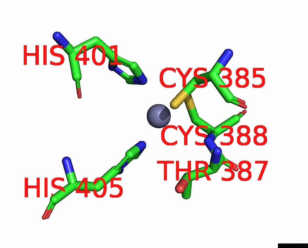
Zinc binding site 1 out of 4 in 2prt
Go back to
Zinc binding site 1 out
of 4 in the Structure of the Wilms Tumor Suppressor Protein Zinc Finger Domain Bound to Dna
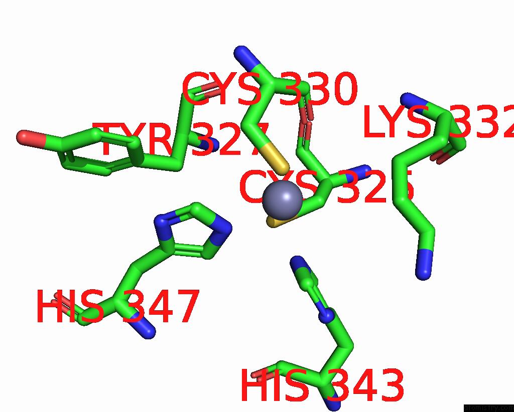
Mono view
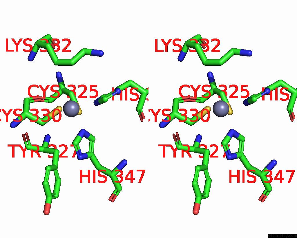
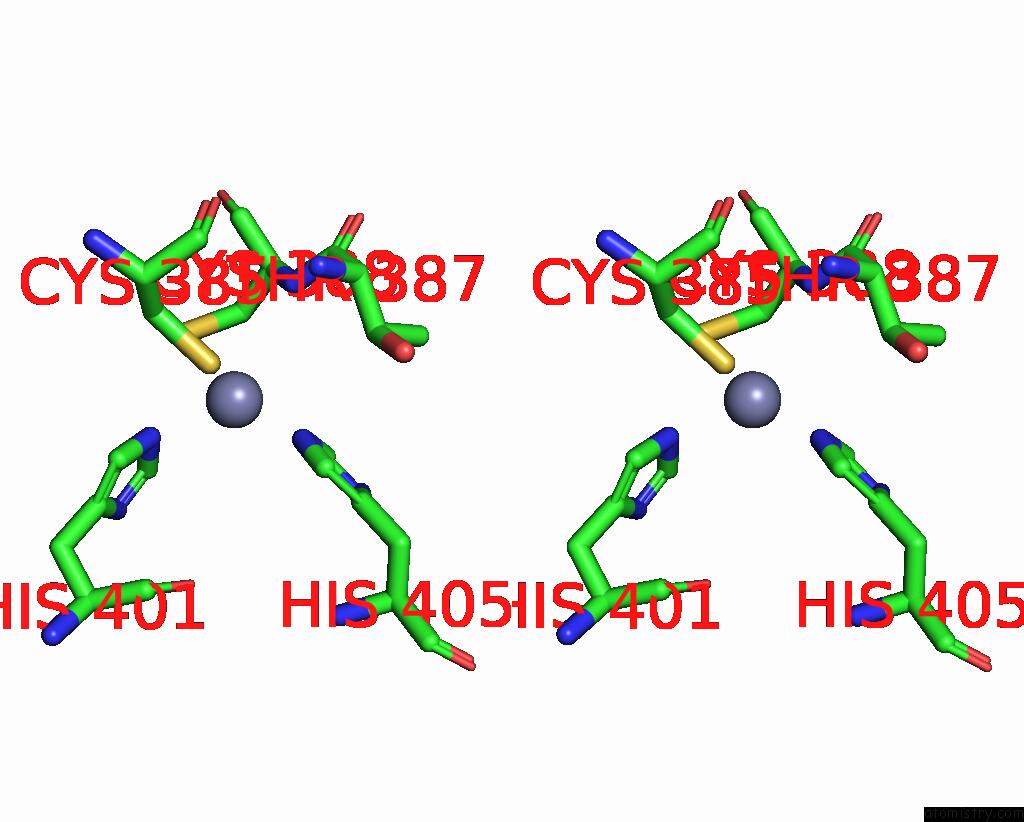
Stereo pair view
Mono view
Stereo pair view
A full contact list of Zinc with other atoms in the Zn binding
site number 1 of Structure of the Wilms Tumor Suppressor Protein Zinc Finger Domain Bound to Dna within 5.0Å range:
|
Zinc binding site 2 out of 4 in 2prt
Go back to
Zinc binding site 2 out
of 4 in the Structure of the Wilms Tumor Suppressor Protein Zinc Finger Domain Bound to Dna
Mono view
Stereo pair view
Mono view
Stereo pair view
A full contact list of Zinc with other atoms in the Zn binding
site number 2 of Structure of the Wilms Tumor Suppressor Protein Zinc Finger Domain Bound to Dna within 5.0Å range:
|
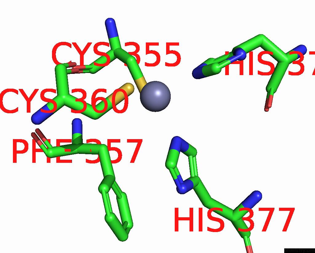
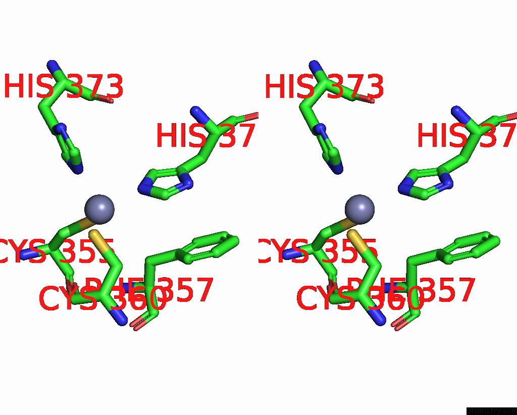
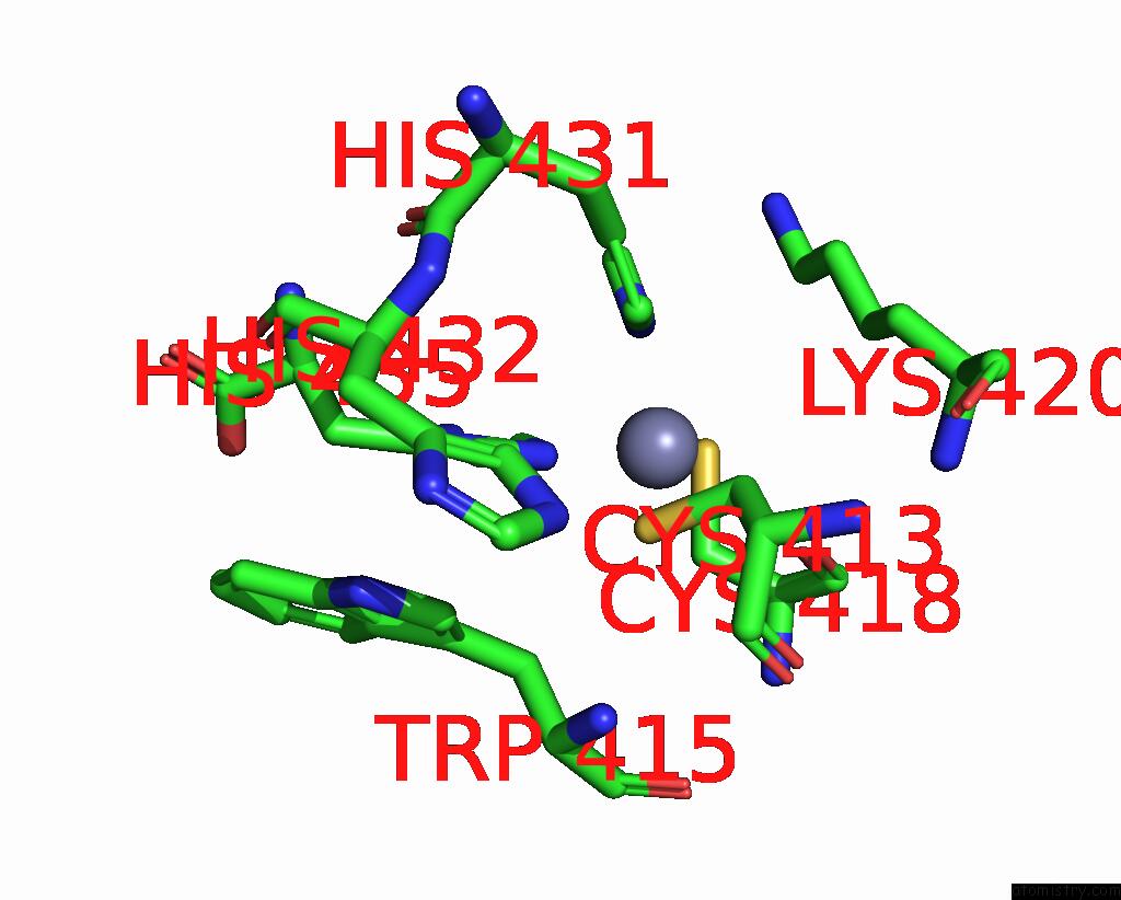
Zinc binding site 3 out of 4 in 2prt
Go back to
Zinc binding site 3 out
of 4 in the Structure of the Wilms Tumor Suppressor Protein Zinc Finger Domain Bound to Dna
Mono view
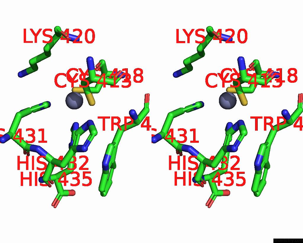
Stereo pair view
Mono view
Stereo pair view
A full contact list of Zinc with other atoms in the Zn binding
site number 3 of Structure of the Wilms Tumor Suppressor Protein Zinc Finger Domain Bound to Dna within 5.0Å range:
|
Zinc binding site 4 out of 4 in 2prt
Go back to
Zinc binding site 4 out
of 4 in the Structure of the Wilms Tumor Suppressor Protein Zinc Finger Domain Bound to Dna
Mono view
Stereo pair view
Mono view
Stereo pair view
A full contact list of Zinc with other atoms in the Zn binding
site number 4 of Structure of the Wilms Tumor Suppressor Protein Zinc Finger Domain Bound to Dna within 5.0Å range:
|
Reference:
R.Stoll,
B.M.Lee,
E.W.Debler,
J.H.Laity,
I.A.Wilson,
H.J.Dyson,
P.E.Wright.
Structure of the Wilms Tumor Suppressor Protein Zinc Finger Domain Bound to Dna J.Mol.Biol. V. 372 1227 2007.
ISSN: ISSN 0022-2836
PubMed: 17716689
DOI: 10.1016/J.JMB.2007.07.017
Page generated: Thu Oct 17 03:11:56 2024
ISSN: ISSN 0022-2836
PubMed: 17716689
DOI: 10.1016/J.JMB.2007.07.017
Last articles
Zn in 9MJ5Zn in 9HNW
Zn in 9G0L
Zn in 9FNE
Zn in 9DZN
Zn in 9E0I
Zn in 9D32
Zn in 9DAK
Zn in 8ZXC
Zn in 8ZUF