Zinc »
PDB 2omi-2ovz »
2ouv »
Zinc in PDB 2ouv: Crystal Structure of PDE10A2 Mutant of D564N
Enzymatic activity of Crystal Structure of PDE10A2 Mutant of D564N
All present enzymatic activity of Crystal Structure of PDE10A2 Mutant of D564N:
3.1.4.17;
3.1.4.17;
Protein crystallography data
The structure of Crystal Structure of PDE10A2 Mutant of D564N, PDB code: 2ouv
was solved by
H.C.Wang,
Y.D.Liu,
J.Hou,
M.Y.Zheng,
H.Robinson,
with X-Ray Crystallography technique. A brief refinement statistics is given in the table below:
| Resolution Low / High (Å) | 30.00 / 1.56 |
| Space group | P 21 21 21 |
| Cell size a, b, c (Å), α, β, γ (°) | 51.441, 82.205, 155.242, 90.00, 90.00, 90.00 |
| R / Rfree (%) | 20.9 / 22.8 |
Other elements in 2ouv:
The structure of Crystal Structure of PDE10A2 Mutant of D564N also contains other interesting chemical elements:
| Magnesium | (Mg) | 2 atoms |
Zinc Binding Sites:
The binding sites of Zinc atom in the Crystal Structure of PDE10A2 Mutant of D564N
(pdb code 2ouv). This binding sites where shown within
5.0 Angstroms radius around Zinc atom.
In total 2 binding sites of Zinc where determined in the Crystal Structure of PDE10A2 Mutant of D564N, PDB code: 2ouv:
Jump to Zinc binding site number: 1; 2;
In total 2 binding sites of Zinc where determined in the Crystal Structure of PDE10A2 Mutant of D564N, PDB code: 2ouv:
Jump to Zinc binding site number: 1; 2;
Zinc binding site 1 out of 2 in 2ouv
Go back to
Zinc binding site 1 out
of 2 in the Crystal Structure of PDE10A2 Mutant of D564N
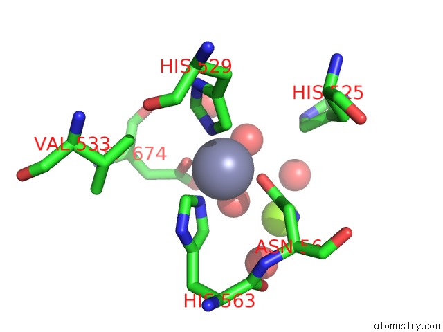
Mono view
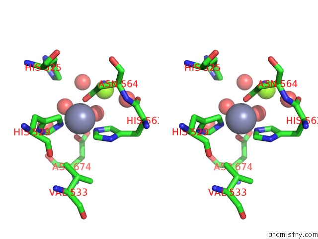
Stereo pair view
Mono view
Stereo pair view
A full contact list of Zinc with other atoms in the Zn binding
site number 1 of Crystal Structure of PDE10A2 Mutant of D564N within 5.0Å range:
|
Zinc binding site 2 out of 2 in 2ouv
Go back to
Zinc binding site 2 out
of 2 in the Crystal Structure of PDE10A2 Mutant of D564N
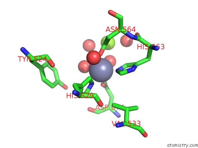
Mono view
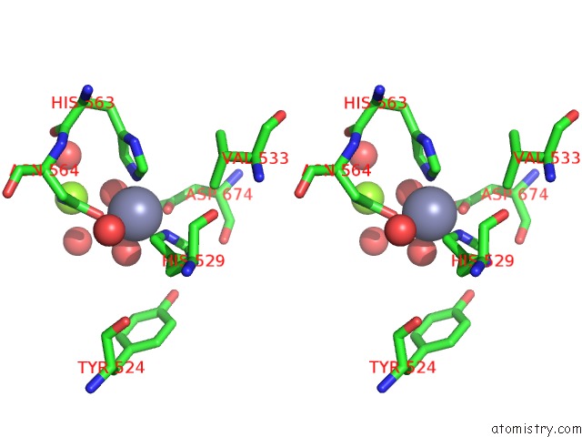
Stereo pair view
Mono view
Stereo pair view
A full contact list of Zinc with other atoms in the Zn binding
site number 2 of Crystal Structure of PDE10A2 Mutant of D564N within 5.0Å range:
|
Reference:
H.Wang,
Y.Liu,
J.Hou,
M.Zheng,
H.Robinson,
H.Ke.
From the Cover: Structural Insight Into Substrate Specificity of Phosphodiesterase 10. Proc.Natl.Acad.Sci.Usa V. 104 5782 2007.
ISSN: ISSN 0027-8424
PubMed: 17389385
DOI: 10.1073/PNAS.0700279104
Page generated: Thu Oct 17 02:47:46 2024
ISSN: ISSN 0027-8424
PubMed: 17389385
DOI: 10.1073/PNAS.0700279104
Last articles
Zn in 9MJ5Zn in 9HNW
Zn in 9G0L
Zn in 9FNE
Zn in 9DZN
Zn in 9E0I
Zn in 9D32
Zn in 9DAK
Zn in 8ZXC
Zn in 8ZUF