Zinc »
PDB 2o8h-2omh »
2om1 »
Zinc in PDB 2om1: Structure of Human Insulin in Presence of Thiocyanate at pH 6.5
Protein crystallography data
The structure of Structure of Human Insulin in Presence of Thiocyanate at pH 6.5, PDB code: 2om1
was solved by
M.Norrman,
G.Schluckebier,
with X-Ray Crystallography technique. A brief refinement statistics is given in the table below:
| Resolution Low / High (Å) | 19.99 / 1.97 |
| Space group | C 2 2 21 1 |
| Cell size a, b, c (Å), α, β, γ (°) | 59.000, 219.480, 224.480, 90.00, 90.00, 90.00 |
| R / Rfree (%) | 17.5 / 21.2 |
Zinc Binding Sites:
The binding sites of Zinc atom in the Structure of Human Insulin in Presence of Thiocyanate at pH 6.5
(pdb code 2om1). This binding sites where shown within
5.0 Angstroms radius around Zinc atom.
In total 6 binding sites of Zinc where determined in the Structure of Human Insulin in Presence of Thiocyanate at pH 6.5, PDB code: 2om1:
Jump to Zinc binding site number: 1; 2; 3; 4; 5; 6;
In total 6 binding sites of Zinc where determined in the Structure of Human Insulin in Presence of Thiocyanate at pH 6.5, PDB code: 2om1:
Jump to Zinc binding site number: 1; 2; 3; 4; 5; 6;
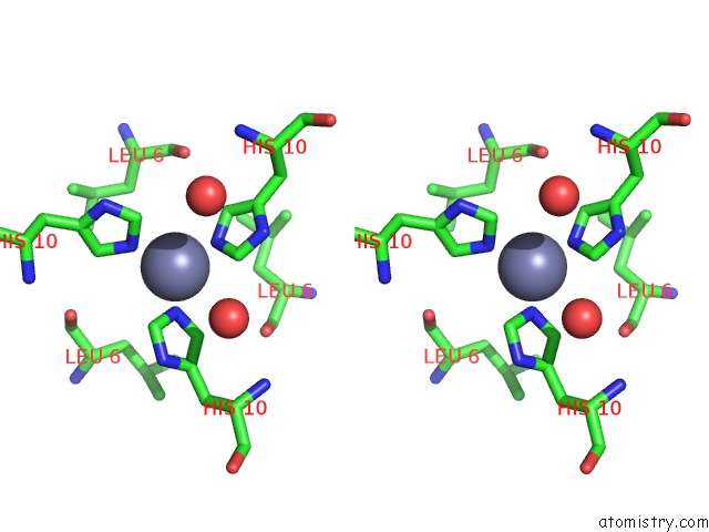
Zinc binding site 1 out of 6 in 2om1
Go back to
Zinc binding site 1 out
of 6 in the Structure of Human Insulin in Presence of Thiocyanate at pH 6.5
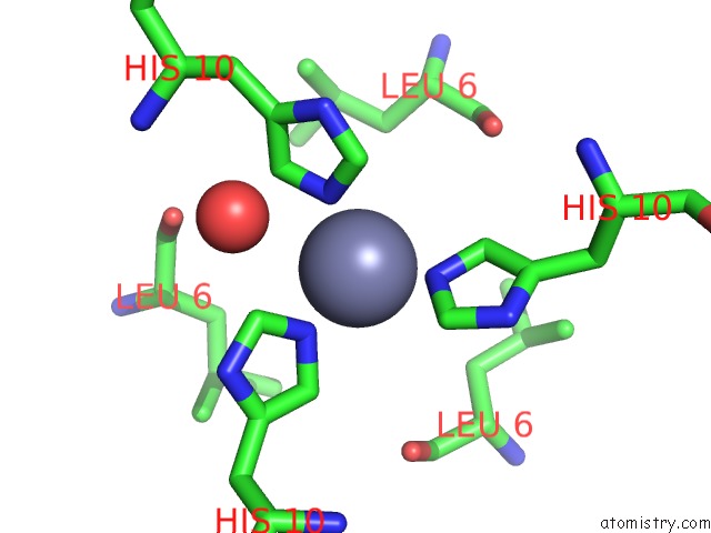
Mono view

Stereo pair view
Mono view
Stereo pair view
A full contact list of Zinc with other atoms in the Zn binding
site number 1 of Structure of Human Insulin in Presence of Thiocyanate at pH 6.5 within 5.0Å range:
|
Zinc binding site 2 out of 6 in 2om1
Go back to
Zinc binding site 2 out
of 6 in the Structure of Human Insulin in Presence of Thiocyanate at pH 6.5
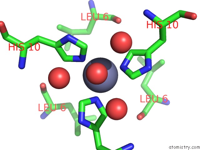
Mono view

Stereo pair view
Mono view
Stereo pair view
A full contact list of Zinc with other atoms in the Zn binding
site number 2 of Structure of Human Insulin in Presence of Thiocyanate at pH 6.5 within 5.0Å range:
|
Zinc binding site 3 out of 6 in 2om1
Go back to
Zinc binding site 3 out
of 6 in the Structure of Human Insulin in Presence of Thiocyanate at pH 6.5
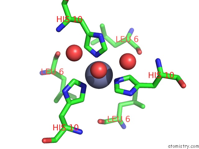
Mono view

Stereo pair view
Mono view
Stereo pair view
A full contact list of Zinc with other atoms in the Zn binding
site number 3 of Structure of Human Insulin in Presence of Thiocyanate at pH 6.5 within 5.0Å range:
|
Zinc binding site 4 out of 6 in 2om1
Go back to
Zinc binding site 4 out
of 6 in the Structure of Human Insulin in Presence of Thiocyanate at pH 6.5
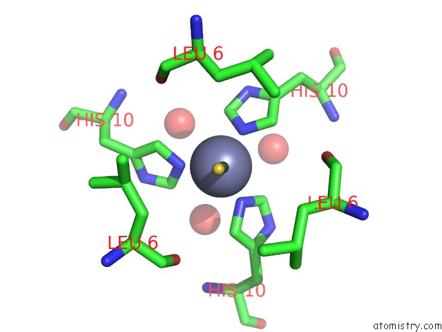
Mono view

Stereo pair view
Mono view
Stereo pair view
A full contact list of Zinc with other atoms in the Zn binding
site number 4 of Structure of Human Insulin in Presence of Thiocyanate at pH 6.5 within 5.0Å range:
|
Zinc binding site 5 out of 6 in 2om1
Go back to
Zinc binding site 5 out
of 6 in the Structure of Human Insulin in Presence of Thiocyanate at pH 6.5

Mono view

Stereo pair view
Mono view
Stereo pair view
A full contact list of Zinc with other atoms in the Zn binding
site number 5 of Structure of Human Insulin in Presence of Thiocyanate at pH 6.5 within 5.0Å range:
|
Zinc binding site 6 out of 6 in 2om1
Go back to
Zinc binding site 6 out
of 6 in the Structure of Human Insulin in Presence of Thiocyanate at pH 6.5

Mono view
Stereo pair view
Mono view
Stereo pair view
A full contact list of Zinc with other atoms in the Zn binding
site number 6 of Structure of Human Insulin in Presence of Thiocyanate at pH 6.5 within 5.0Å range:
|
Reference:
M.Norrman,
G.Schluckebier.
Crystallographic Characterization of Two Novel Crystal Forms of Human Insulin Induced By Chaotropic Agents and A Shift in pH. Bmc Struct.Biol. V. 7 83 2007.
ISSN: ESSN 1472-6807
PubMed: 18093308
DOI: 10.1186/1472-6807-7-83
Page generated: Thu Oct 17 02:40:46 2024
ISSN: ESSN 1472-6807
PubMed: 18093308
DOI: 10.1186/1472-6807-7-83
Last articles
Al in 7NVOAl in 7NIC
Al in 7NIQ
Al in 7L07
Al in 7N77
Al in 7N73
Al in 7N72
Al in 7LVR
Al in 7KYB
Al in 7JL3