Zinc »
PDB 2o8j-2omi »
2oh3 »
Zinc in PDB 2oh3: Crystal Structure of COG1633: Uncharacterized Conserved Protein (ZP_00055496.1) From Magnetospirillum Magnetotacticum Ms-1 at 2.00 A Resolution
Protein crystallography data
The structure of Crystal Structure of COG1633: Uncharacterized Conserved Protein (ZP_00055496.1) From Magnetospirillum Magnetotacticum Ms-1 at 2.00 A Resolution, PDB code: 2oh3
was solved by
Joint Center For Structural Genomics (Jcsg),
with X-Ray Crystallography technique. A brief refinement statistics is given in the table below:
| Resolution Low / High (Å) | 28.11 / 2.00 |
| Space group | I 2 2 2 |
| Cell size a, b, c (Å), α, β, γ (°) | 64.360, 75.280, 89.790, 90.00, 90.00, 90.00 |
| R / Rfree (%) | 19.2 / 23.5 |
Zinc Binding Sites:
The binding sites of Zinc atom in the Crystal Structure of COG1633: Uncharacterized Conserved Protein (ZP_00055496.1) From Magnetospirillum Magnetotacticum Ms-1 at 2.00 A Resolution
(pdb code 2oh3). This binding sites where shown within
5.0 Angstroms radius around Zinc atom.
In total only one binding site of Zinc was determined in the Crystal Structure of COG1633: Uncharacterized Conserved Protein (ZP_00055496.1) From Magnetospirillum Magnetotacticum Ms-1 at 2.00 A Resolution, PDB code: 2oh3:
In total only one binding site of Zinc was determined in the Crystal Structure of COG1633: Uncharacterized Conserved Protein (ZP_00055496.1) From Magnetospirillum Magnetotacticum Ms-1 at 2.00 A Resolution, PDB code: 2oh3:
Zinc binding site 1 out of 1 in 2oh3
Go back to
Zinc binding site 1 out
of 1 in the Crystal Structure of COG1633: Uncharacterized Conserved Protein (ZP_00055496.1) From Magnetospirillum Magnetotacticum Ms-1 at 2.00 A Resolution
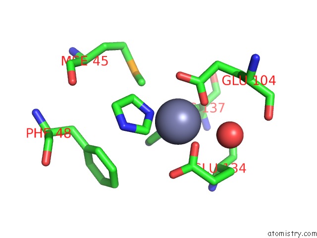
Mono view
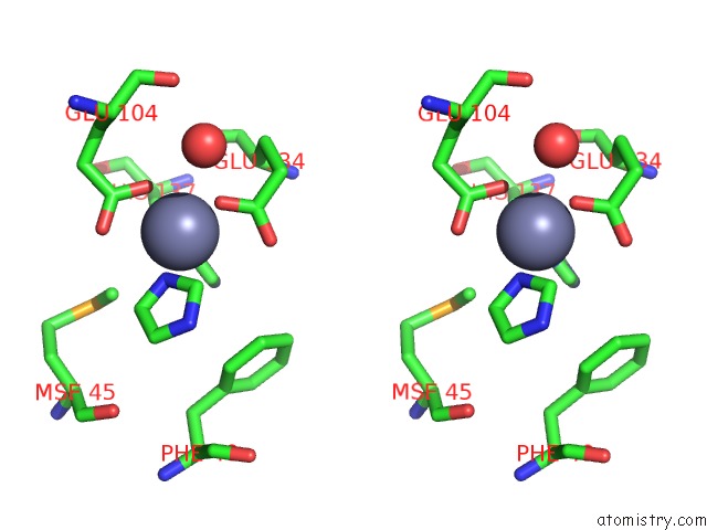
Stereo pair view
Mono view
Stereo pair view
A full contact list of Zinc with other atoms in the Zn binding
site number 1 of Crystal Structure of COG1633: Uncharacterized Conserved Protein (ZP_00055496.1) From Magnetospirillum Magnetotacticum Ms-1 at 2.00 A Resolution within 5.0Å range:
|
Reference:
Joint Center For Structural Genomics (Jcsg),
Joint Center For Structural Genomics (Jcsg).
N/A N/A.
Page generated: Wed Aug 20 04:49:32 2025
Last articles
Zn in 3FEQZn in 3FKI
Zn in 3FL2
Zn in 3FLF
Zn in 3FJU
Zn in 3FKG
Zn in 3FID
Zn in 3FII
Zn in 3FIG
Zn in 3FIE