Zinc »
PDB 2o8h-2omh »
2ogw »
Zinc in PDB 2ogw: Structure of Abc Type Zinc Transporter From E. Coli
Protein crystallography data
The structure of Structure of Abc Type Zinc Transporter From E. Coli, PDB code: 2ogw
was solved by
A.Sharma,
M.Yogavel,
with X-Ray Crystallography technique. A brief refinement statistics is given in the table below:
| Resolution Low / High (Å) | 61.66 / 1.83 |
| Space group | P 21 21 21 |
| Cell size a, b, c (Å), α, β, γ (°) | 72.900, 86.378, 87.990, 90.00, 90.00, 90.00 |
| R / Rfree (%) | 22.3 / 26.9 |
Zinc Binding Sites:
The binding sites of Zinc atom in the Structure of Abc Type Zinc Transporter From E. Coli
(pdb code 2ogw). This binding sites where shown within
5.0 Angstroms radius around Zinc atom.
In total 2 binding sites of Zinc where determined in the Structure of Abc Type Zinc Transporter From E. Coli, PDB code: 2ogw:
Jump to Zinc binding site number: 1; 2;
In total 2 binding sites of Zinc where determined in the Structure of Abc Type Zinc Transporter From E. Coli, PDB code: 2ogw:
Jump to Zinc binding site number: 1; 2;
Zinc binding site 1 out of 2 in 2ogw
Go back to
Zinc binding site 1 out
of 2 in the Structure of Abc Type Zinc Transporter From E. Coli

Mono view
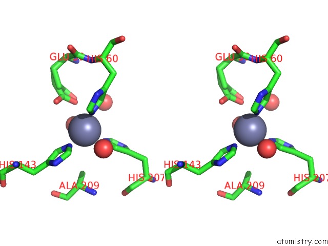
Stereo pair view
Mono view
Stereo pair view
A full contact list of Zinc with other atoms in the Zn binding
site number 1 of Structure of Abc Type Zinc Transporter From E. Coli within 5.0Å range:
|
Zinc binding site 2 out of 2 in 2ogw
Go back to
Zinc binding site 2 out
of 2 in the Structure of Abc Type Zinc Transporter From E. Coli

Mono view
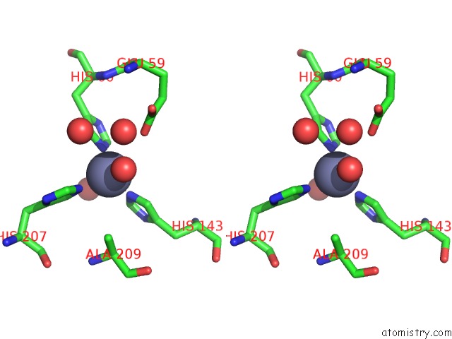
Stereo pair view
Mono view
Stereo pair view
A full contact list of Zinc with other atoms in the Zn binding
site number 2 of Structure of Abc Type Zinc Transporter From E. Coli within 5.0Å range:
|
Reference:
B.R.Chandra,
M.Yogavel,
A.Sharma.
Structural Analysis of Abc-Family Periplasmic Zinc Binding Protein Provides New Insights Into Mechanism of Ligand Uptake and Release J.Mol.Biol. V. 367 970 2007.
ISSN: ISSN 0022-2836
PubMed: 17306297
DOI: 10.1016/J.JMB.2007.01.041
Page generated: Thu Oct 17 02:38:05 2024
ISSN: ISSN 0022-2836
PubMed: 17306297
DOI: 10.1016/J.JMB.2007.01.041
Last articles
Al in 8SHDAl in 8SHA
Al in 8SGL
Al in 8SH9
Al in 8SGC
Al in 8SG9
Al in 8SFF
Al in 8SG8
Al in 8R1A
Al in 8Q75