Zinc »
PDB 2o8h-2omh »
2od7 »
Zinc in PDB 2od7: Crystal Structure of YHST2 Bound to the Intermediate Analogue Adp-Hpd, and and Aceylated H4 Peptide
Protein crystallography data
The structure of Crystal Structure of YHST2 Bound to the Intermediate Analogue Adp-Hpd, and and Aceylated H4 Peptide, PDB code: 2od7
was solved by
R.Q.Marmorstein,
B.D.Sanders,
with X-Ray Crystallography technique. A brief refinement statistics is given in the table below:
| Resolution Low / High (Å) | 41.92 / 2.00 |
| Space group | P 32 2 1 |
| Cell size a, b, c (Å), α, β, γ (°) | 106.759, 106.759, 67.689, 90.00, 90.00, 120.00 |
| R / Rfree (%) | 22.2 / 23.4 |
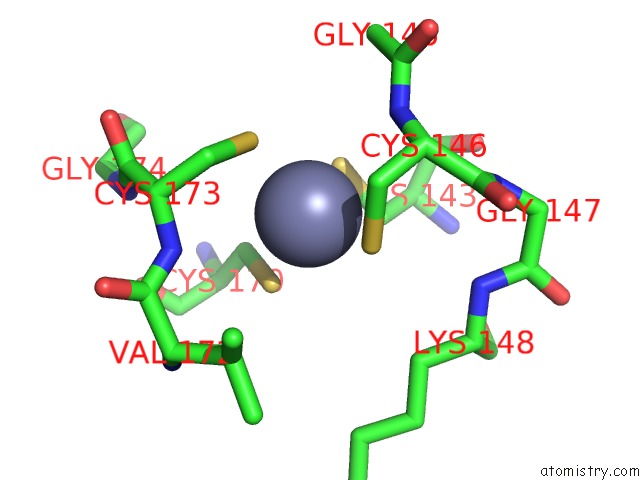
Zinc Binding Sites:
The binding sites of Zinc atom in the Crystal Structure of YHST2 Bound to the Intermediate Analogue Adp-Hpd, and and Aceylated H4 Peptide
(pdb code 2od7). This binding sites where shown within
5.0 Angstroms radius around Zinc atom.
In total only one binding site of Zinc was determined in the Crystal Structure of YHST2 Bound to the Intermediate Analogue Adp-Hpd, and and Aceylated H4 Peptide, PDB code: 2od7:
In total only one binding site of Zinc was determined in the Crystal Structure of YHST2 Bound to the Intermediate Analogue Adp-Hpd, and and Aceylated H4 Peptide, PDB code: 2od7:
Zinc binding site 1 out of 1 in 2od7
Go back to
Zinc binding site 1 out
of 1 in the Crystal Structure of YHST2 Bound to the Intermediate Analogue Adp-Hpd, and and Aceylated H4 Peptide
Mono view

Stereo pair view
Mono view
Stereo pair view
A full contact list of Zinc with other atoms in the Zn binding
site number 1 of Crystal Structure of YHST2 Bound to the Intermediate Analogue Adp-Hpd, and and Aceylated H4 Peptide within 5.0Å range:
|
Reference:
B.D.Sanders,
K.Zhao,
J.T.Slama,
R.Marmorstein.
Structural Basis For Nicotinamide Inhibition and Base Exchange in SIR2 Enzymes. Mol.Cell V. 25 463 2007.
ISSN: ISSN 1097-2765
PubMed: 17289592
DOI: 10.1016/J.MOLCEL.2006.12.022
Page generated: Thu Oct 17 02:37:09 2024
ISSN: ISSN 1097-2765
PubMed: 17289592
DOI: 10.1016/J.MOLCEL.2006.12.022
Last articles
Zn in 9MJ5Zn in 9HNW
Zn in 9G0L
Zn in 9FNE
Zn in 9DZN
Zn in 9E0I
Zn in 9D32
Zn in 9DAK
Zn in 8ZXC
Zn in 8ZUF