Zinc in PDB 2od1: Solution Structure of the Mynd Domain From Human AML1-Eto
Zinc Binding Sites:
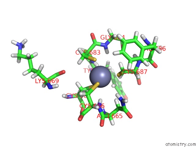
The binding sites of Zinc atom in the Solution Structure of the Mynd Domain From Human AML1-Eto
(pdb code 2od1). This binding sites where shown within
5.0 Angstroms radius around Zinc atom.
In total 2 binding sites of Zinc where determined in the
Solution Structure of the Mynd Domain From Human AML1-Eto, PDB code: 2od1:
Jump to Zinc binding site number:
1;
2;
Zinc binding site 1 out
of 2 in 2od1
Go back to
Zinc Binding Sites List in 2od1
Zinc binding site 1 out
of 2 in the Solution Structure of the Mynd Domain From Human AML1-Eto
Mono view
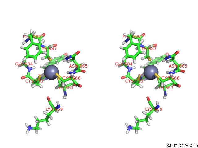
Stereo pair view
|
A full contact list of Zinc with other atoms in the Zn binding
site number 1 of Solution Structure of the Mynd Domain From Human AML1-Eto within 5.0Å range:
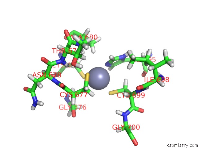
probe
|
atom
|
residue
|
distance (Å)
|
B
|
Occ
|
A:Zn901
b:0.0
occ:1.00
|
SG
|
A:CYS663
|
2.3
|
0.0
|
1.0
|
SG
|
A:CYS683
|
2.3
|
0.0
|
1.0
|
SG
|
A:CYS666
|
2.3
|
0.0
|
1.0
|
SG
|
A:CYS687
|
2.3
|
0.0
|
1.0
|
HB2
|
A:CYS666
|
2.9
|
0.0
|
1.0
|
H
|
A:CYS683
|
2.9
|
0.0
|
1.0
|
H
|
A:CYS666
|
3.0
|
0.0
|
1.0
|
CB
|
A:CYS666
|
3.2
|
0.0
|
1.0
|
HD21
|
A:ASN665
|
3.3
|
0.0
|
1.0
|
CB
|
A:CYS663
|
3.4
|
0.0
|
1.0
|
HB3
|
A:CYS683
|
3.4
|
0.0
|
1.0
|
HB2
|
A:CYS663
|
3.4
|
0.0
|
1.0
|
CB
|
A:CYS687
|
3.4
|
0.0
|
1.0
|
HB3
|
A:CYS687
|
3.5
|
0.0
|
1.0
|
CB
|
A:CYS683
|
3.5
|
0.0
|
1.0
|
HB3
|
A:CYS663
|
3.5
|
0.0
|
1.0
|
HB2
|
A:CYS687
|
3.6
|
0.0
|
1.0
|
N
|
A:CYS683
|
3.8
|
0.0
|
1.0
|
N
|
A:CYS666
|
3.8
|
0.0
|
1.0
|
HB3
|
A:ASN665
|
3.8
|
0.0
|
1.0
|
HB3
|
A:CYS666
|
4.0
|
0.0
|
1.0
|
CA
|
A:CYS666
|
4.1
|
0.0
|
1.0
|
CA
|
A:CYS683
|
4.2
|
0.0
|
1.0
|
H
|
A:GLY684
|
4.2
|
0.0
|
1.0
|
HZ
|
A:PHE686
|
4.2
|
0.0
|
1.0
|
ND2
|
A:ASN665
|
4.2
|
0.0
|
1.0
|
HB3
|
A:TYR682
|
4.3
|
0.0
|
1.0
|
HB2
|
A:CYS683
|
4.3
|
0.0
|
1.0
|
HD1
|
A:TYR682
|
4.4
|
0.0
|
1.0
|
HA
|
A:TYR682
|
4.4
|
0.0
|
1.0
|
H
|
A:ASN665
|
4.4
|
0.0
|
1.0
|
HE2
|
A:PHE686
|
4.5
|
0.0
|
1.0
|
HA
|
A:CYS666
|
4.5
|
0.0
|
1.0
|
CZ
|
A:PHE686
|
4.6
|
0.0
|
1.0
|
N
|
A:GLY684
|
4.7
|
0.0
|
1.0
|
CB
|
A:ASN665
|
4.8
|
0.0
|
1.0
|
CE2
|
A:PHE686
|
4.8
|
0.0
|
1.0
|
CA
|
A:CYS663
|
4.8
|
0.0
|
1.0
|
HD22
|
A:ASN665
|
4.8
|
0.0
|
1.0
|
C
|
A:CYS683
|
4.8
|
0.0
|
1.0
|
O
|
A:LYS669
|
4.8
|
0.0
|
1.0
|
C
|
A:TYR682
|
4.8
|
0.0
|
1.0
|
CA
|
A:CYS687
|
4.8
|
0.0
|
1.0
|
HA
|
A:CYS687
|
5.0
|
0.0
|
1.0
|
C
|
A:ASN665
|
5.0
|
0.0
|
1.0
|
|
Zinc binding site 2 out
of 2 in 2od1
Go back to
Zinc Binding Sites List in 2od1
Zinc binding site 2 out
of 2 in the Solution Structure of the Mynd Domain From Human AML1-Eto
Mono view
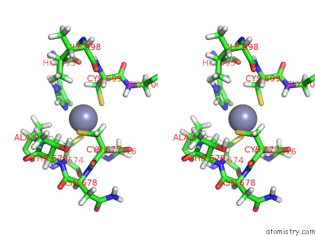
Stereo pair view
|
A full contact list of Zinc with other atoms in the Zn binding
site number 2 of Solution Structure of the Mynd Domain From Human AML1-Eto within 5.0Å range:
probe
|
atom
|
residue
|
distance (Å)
|
B
|
Occ
|
A:Zn902
b:0.0
occ:1.00
|
NE2
|
A:HIS695
|
2.0
|
0.0
|
1.0
|
SG
|
A:CYS674
|
2.3
|
0.0
|
1.0
|
SG
|
A:CYS699
|
2.3
|
0.0
|
1.0
|
SG
|
A:CYS677
|
2.3
|
0.0
|
1.0
|
CD2
|
A:HIS695
|
2.9
|
0.0
|
1.0
|
CE1
|
A:HIS695
|
3.0
|
0.0
|
1.0
|
HB3
|
A:CYS677
|
3.0
|
0.0
|
1.0
|
HD2
|
A:HIS695
|
3.1
|
0.0
|
1.0
|
CB
|
A:CYS677
|
3.2
|
0.0
|
1.0
|
HE1
|
A:HIS695
|
3.3
|
0.0
|
1.0
|
HB3
|
A:ALA680
|
3.3
|
0.0
|
1.0
|
HB2
|
A:CYS699
|
3.3
|
0.0
|
1.0
|
CB
|
A:CYS674
|
3.4
|
0.0
|
1.0
|
HB3
|
A:CYS674
|
3.4
|
0.0
|
1.0
|
H
|
A:CYS677
|
3.4
|
0.0
|
1.0
|
HB2
|
A:CYS674
|
3.5
|
0.0
|
1.0
|
CB
|
A:CYS699
|
3.5
|
0.0
|
1.0
|
N
|
A:CYS677
|
3.8
|
0.0
|
1.0
|
CG
|
A:HIS695
|
4.0
|
0.0
|
1.0
|
ND1
|
A:HIS695
|
4.0
|
0.0
|
1.0
|
H
|
A:ALA680
|
4.0
|
0.0
|
1.0
|
HB2
|
A:CYS677
|
4.1
|
0.0
|
1.0
|
CA
|
A:CYS677
|
4.1
|
0.0
|
1.0
|
HB3
|
A:CYS699
|
4.2
|
0.0
|
1.0
|
CB
|
A:ALA680
|
4.3
|
0.0
|
1.0
|
HA
|
A:CYS699
|
4.3
|
0.0
|
1.0
|
HA3
|
A:GLY676
|
4.4
|
0.0
|
1.0
|
H
|
A:GLY676
|
4.5
|
0.0
|
1.0
|
C
|
A:GLY676
|
4.5
|
0.0
|
1.0
|
HB2
|
A:ALA680
|
4.5
|
0.0
|
1.0
|
CA
|
A:CYS699
|
4.6
|
0.0
|
1.0
|
H
|
A:THR679
|
4.6
|
0.0
|
1.0
|
HB
|
A:THR679
|
4.8
|
0.0
|
1.0
|
CA
|
A:CYS674
|
4.8
|
0.0
|
1.0
|
HB1
|
A:ALA680
|
4.8
|
0.0
|
1.0
|
H
|
A:ASN678
|
4.8
|
0.0
|
1.0
|
HD1
|
A:HIS695
|
4.9
|
0.0
|
1.0
|
CA
|
A:GLY676
|
4.9
|
0.0
|
1.0
|
N
|
A:ALA680
|
4.9
|
0.0
|
1.0
|
HD13
|
A:ILE698
|
4.9
|
0.0
|
1.0
|
HA
|
A:CYS677
|
4.9
|
0.0
|
1.0
|
H
|
A:GLY700
|
5.0
|
0.0
|
1.0
|
C
|
A:CYS677
|
5.0
|
0.0
|
1.0
|
|
Reference:
Y.Liu,
W.Chen,
J.Gaudet,
M.D.Cheney,
L.Roudaia,
T.Cierpicki,
R.C.Klet,
K.Hartman,
T.M.Laue,
N.A.Speck,
J.H.Bushweller.
Structural Basis For Recognition of Smrt/N-Cor By the Mynd Domain and Its Contribution to AML1/Eto'S Activity. Cancer Cell V. 11 483 2007.
ISSN: ISSN 1535-6108
PubMed: 17560331
DOI: 10.1016/J.CCR.2007.04.010
Page generated: Thu Oct 17 02:36:23 2024
|