Zinc »
PDB 2nwz-2o6p »
2o6i »
Zinc in PDB 2o6i: Structure of An Enterococcus Faecalis Hd Domain Phosphohydrolase
Protein crystallography data
The structure of Structure of An Enterococcus Faecalis Hd Domain Phosphohydrolase, PDB code: 2o6i
was solved by
I.I.Vorontsov,
G.Minasov,
L.Shuvalova,
J.S.Brunzelle,
S.Moy,
F.R.Collart,
A.Joachimiak,
W.F.Anderson,
Midwest Center For Structural Genomics(Mcsg),
with X-Ray Crystallography technique. A brief refinement statistics is given in the table below:
| Resolution Low / High (Å) | 30.00 / 2.55 |
| Space group | P 32 2 1 |
| Cell size a, b, c (Å), α, β, γ (°) | 109.913, 109.913, 182.412, 90.00, 90.00, 120.00 |
| R / Rfree (%) | 24.8 / 31.7 |
Other elements in 2o6i:
The structure of Structure of An Enterococcus Faecalis Hd Domain Phosphohydrolase also contains other interesting chemical elements:
| Chlorine | (Cl) | 2 atoms |
Zinc Binding Sites:
The binding sites of Zinc atom in the Structure of An Enterococcus Faecalis Hd Domain Phosphohydrolase
(pdb code 2o6i). This binding sites where shown within
5.0 Angstroms radius around Zinc atom.
In total 2 binding sites of Zinc where determined in the Structure of An Enterococcus Faecalis Hd Domain Phosphohydrolase, PDB code: 2o6i:
Jump to Zinc binding site number: 1; 2;
In total 2 binding sites of Zinc where determined in the Structure of An Enterococcus Faecalis Hd Domain Phosphohydrolase, PDB code: 2o6i:
Jump to Zinc binding site number: 1; 2;
Zinc binding site 1 out of 2 in 2o6i
Go back to
Zinc binding site 1 out
of 2 in the Structure of An Enterococcus Faecalis Hd Domain Phosphohydrolase

Mono view
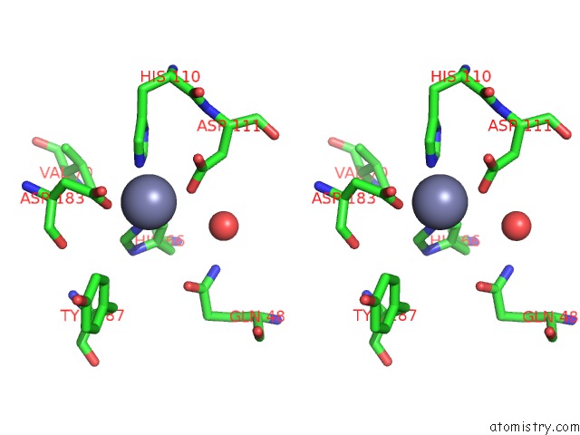
Stereo pair view
Mono view
Stereo pair view
A full contact list of Zinc with other atoms in the Zn binding
site number 1 of Structure of An Enterococcus Faecalis Hd Domain Phosphohydrolase within 5.0Å range:
|
Zinc binding site 2 out of 2 in 2o6i
Go back to
Zinc binding site 2 out
of 2 in the Structure of An Enterococcus Faecalis Hd Domain Phosphohydrolase

Mono view

Stereo pair view
Mono view
Stereo pair view
A full contact list of Zinc with other atoms in the Zn binding
site number 2 of Structure of An Enterococcus Faecalis Hd Domain Phosphohydrolase within 5.0Å range:
|
Reference:
I.I.Vorontsov,
G.Minasov,
O.Kiryukhina,
J.S.Brunzelle,
L.Shuvalova,
W.F.Anderson.
Characterization of the Deoxynucleotide Triphosphate Triphosphohydrolase (Dntpase) Activity of the EF1143 Protein From Enterococcus Faecalis and Crystal Structure of the Activator-Substrate Complex. J.Biol.Chem. V. 286 33158 2011.
ISSN: ISSN 0021-9258
PubMed: 21757692
DOI: 10.1074/JBC.M111.250456
Page generated: Thu Oct 17 02:29:49 2024
ISSN: ISSN 0021-9258
PubMed: 21757692
DOI: 10.1074/JBC.M111.250456
Last articles
Al in 7RE2Al in 7RE0
Al in 7RDX
Al in 7RDY
Al in 7QV9
Al in 7RD7
Al in 7OTJ
Al in 7QBZ
Al in 7OL3
Al in 7OP1