Zinc »
PDB 2nwz-2o6p »
2o4q »
Zinc in PDB 2o4q: Structure of Phosphotriesterase Mutant G60A
Enzymatic activity of Structure of Phosphotriesterase Mutant G60A
All present enzymatic activity of Structure of Phosphotriesterase Mutant G60A:
3.1.8.1;
3.1.8.1;
Protein crystallography data
The structure of Structure of Phosphotriesterase Mutant G60A, PDB code: 2o4q
was solved by
J.Kim,
U.A.Ramagopal,
P.C.Tsai,
F.M.Raushel,
S.C.Almo,
with X-Ray Crystallography technique. A brief refinement statistics is given in the table below:
| Resolution Low / High (Å) | 31.64 / 1.95 |
| Space group | P 1 |
| Cell size a, b, c (Å), α, β, γ (°) | 55.295, 68.299, 90.030, 90.05, 100.42, 89.96 |
| R / Rfree (%) | 16.4 / 22.6 |
Other elements in 2o4q:
The structure of Structure of Phosphotriesterase Mutant G60A also contains other interesting chemical elements:
| Arsenic | (As) | 4 atoms |
Zinc Binding Sites:
The binding sites of Zinc atom in the Structure of Phosphotriesterase Mutant G60A
(pdb code 2o4q). This binding sites where shown within
5.0 Angstroms radius around Zinc atom.
In total 8 binding sites of Zinc where determined in the Structure of Phosphotriesterase Mutant G60A, PDB code: 2o4q:
Jump to Zinc binding site number: 1; 2; 3; 4; 5; 6; 7; 8;
In total 8 binding sites of Zinc where determined in the Structure of Phosphotriesterase Mutant G60A, PDB code: 2o4q:
Jump to Zinc binding site number: 1; 2; 3; 4; 5; 6; 7; 8;
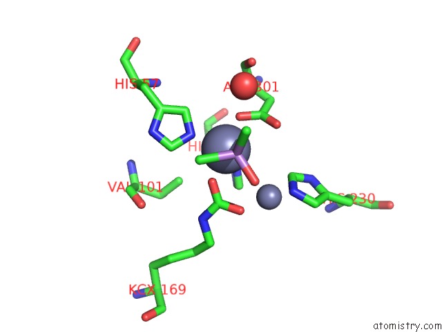
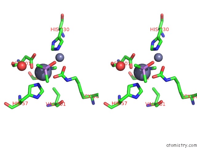
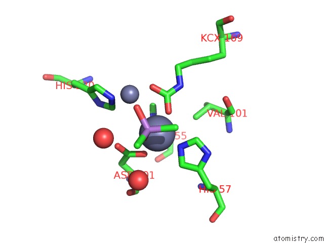
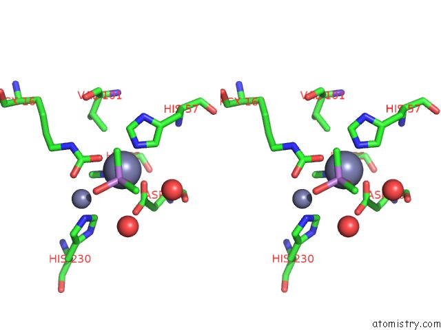
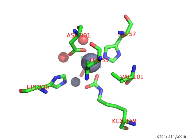
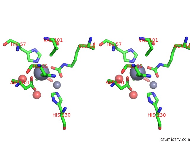
Zinc binding site 1 out of 8 in 2o4q
Go back to
Zinc binding site 1 out
of 8 in the Structure of Phosphotriesterase Mutant G60A
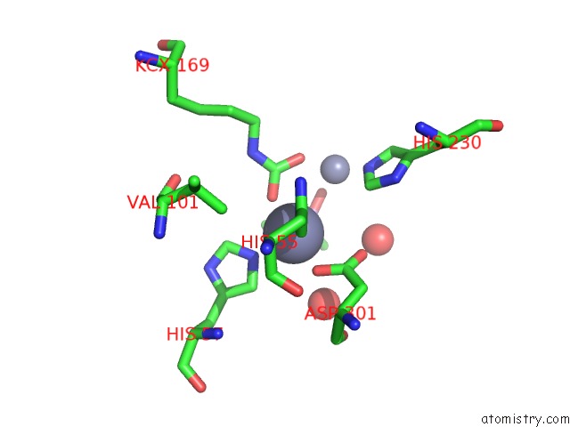
Mono view
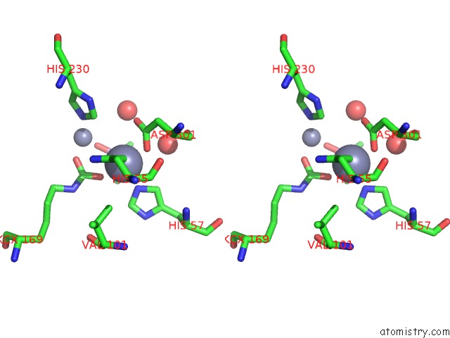
Stereo pair view
Mono view
Stereo pair view
A full contact list of Zinc with other atoms in the Zn binding
site number 1 of Structure of Phosphotriesterase Mutant G60A within 5.0Å range:
|
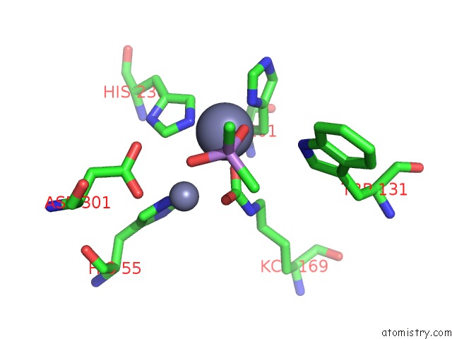
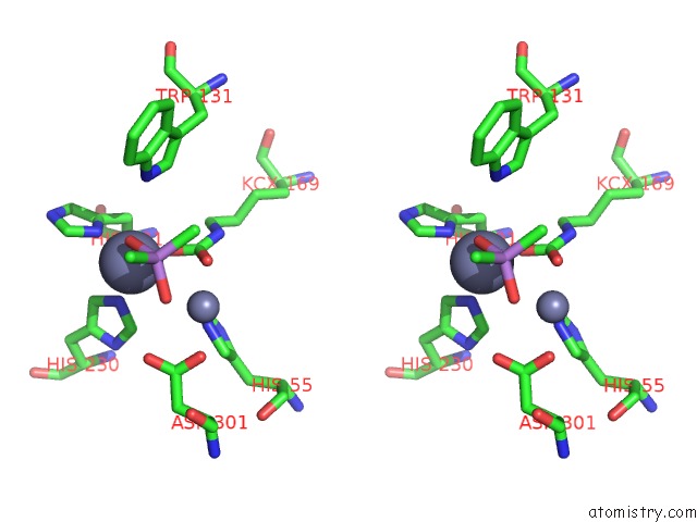
Zinc binding site 2 out of 8 in 2o4q
Go back to
Zinc binding site 2 out
of 8 in the Structure of Phosphotriesterase Mutant G60A
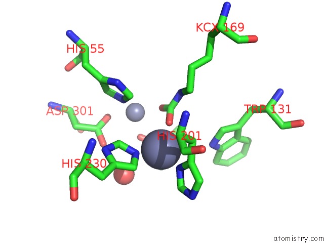
Mono view
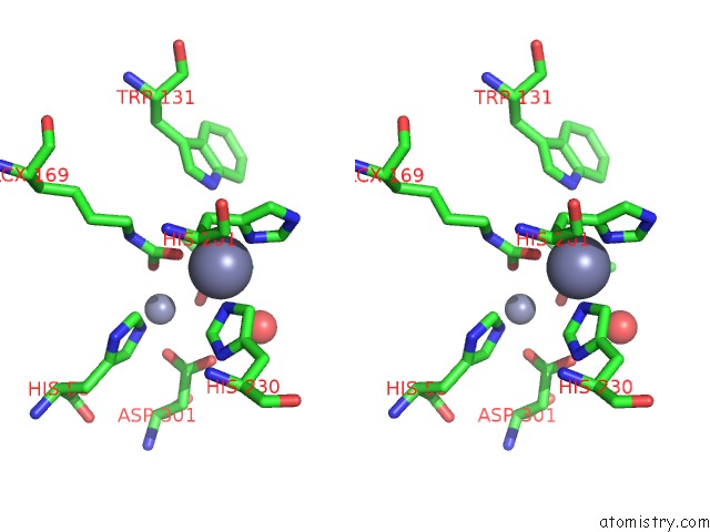
Stereo pair view
Mono view
Stereo pair view
A full contact list of Zinc with other atoms in the Zn binding
site number 2 of Structure of Phosphotriesterase Mutant G60A within 5.0Å range:
|
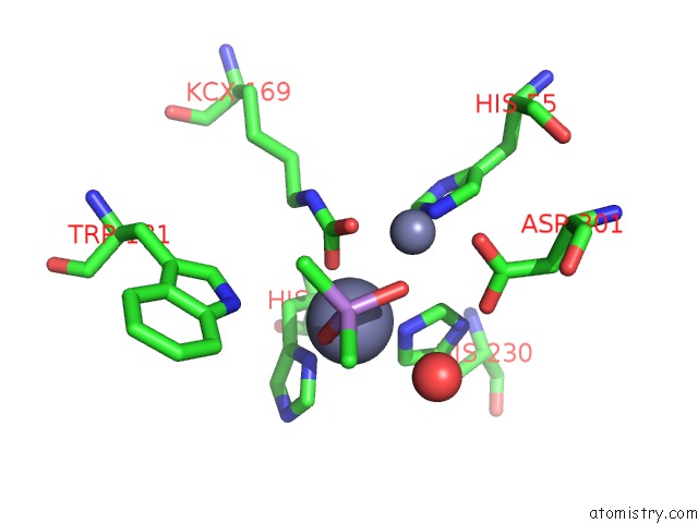
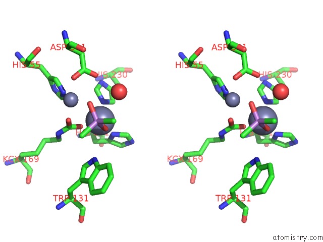
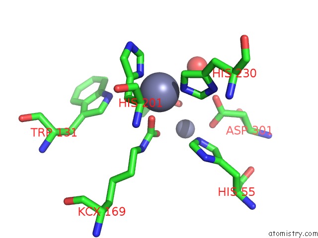
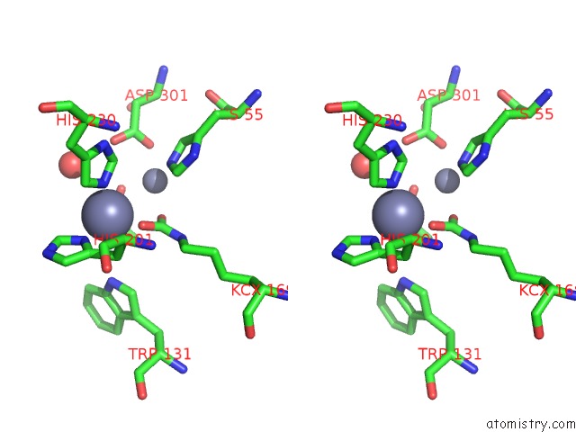
Zinc binding site 3 out of 8 in 2o4q
Go back to
Zinc binding site 3 out
of 8 in the Structure of Phosphotriesterase Mutant G60A
Mono view
Stereo pair view
Mono view
Stereo pair view
A full contact list of Zinc with other atoms in the Zn binding
site number 3 of Structure of Phosphotriesterase Mutant G60A within 5.0Å range:
|
Zinc binding site 4 out of 8 in 2o4q
Go back to
Zinc binding site 4 out
of 8 in the Structure of Phosphotriesterase Mutant G60A
Mono view
Stereo pair view
Mono view
Stereo pair view
A full contact list of Zinc with other atoms in the Zn binding
site number 4 of Structure of Phosphotriesterase Mutant G60A within 5.0Å range:
|
Zinc binding site 5 out of 8 in 2o4q
Go back to
Zinc binding site 5 out
of 8 in the Structure of Phosphotriesterase Mutant G60A
Mono view
Stereo pair view
Mono view
Stereo pair view
A full contact list of Zinc with other atoms in the Zn binding
site number 5 of Structure of Phosphotriesterase Mutant G60A within 5.0Å range:
|
Zinc binding site 6 out of 8 in 2o4q
Go back to
Zinc binding site 6 out
of 8 in the Structure of Phosphotriesterase Mutant G60A
Mono view
Stereo pair view
Mono view
Stereo pair view
A full contact list of Zinc with other atoms in the Zn binding
site number 6 of Structure of Phosphotriesterase Mutant G60A within 5.0Å range:
|
Zinc binding site 7 out of 8 in 2o4q
Go back to
Zinc binding site 7 out
of 8 in the Structure of Phosphotriesterase Mutant G60A
Mono view
Stereo pair view
Mono view
Stereo pair view
A full contact list of Zinc with other atoms in the Zn binding
site number 7 of Structure of Phosphotriesterase Mutant G60A within 5.0Å range:
|
Zinc binding site 8 out of 8 in 2o4q
Go back to
Zinc binding site 8 out
of 8 in the Structure of Phosphotriesterase Mutant G60A
Mono view
Stereo pair view
Mono view
Stereo pair view
A full contact list of Zinc with other atoms in the Zn binding
site number 8 of Structure of Phosphotriesterase Mutant G60A within 5.0Å range:
|
Reference:
J.Kim,
P.C.Tsai,
S.L.Chen,
F.Himo,
S.C.Almo,
F.M.Raushel.
Structure of Diethyl Phosphate Bound to the Binuclear Metal Center of Phosphotriesterase. Biochemistry V. 47 9497 2008.
ISSN: ISSN 0006-2960
PubMed: 18702530
DOI: 10.1021/BI800971V
Page generated: Thu Oct 17 02:26:52 2024
ISSN: ISSN 0006-2960
PubMed: 18702530
DOI: 10.1021/BI800971V
Last articles
Al in 7NICAl in 7NIQ
Al in 7L07
Al in 7N77
Al in 7N73
Al in 7N72
Al in 7LVR
Al in 7KYB
Al in 7JL3
Al in 7KRO