Zinc in PDB 2l0z: Solution Structure of A Zinc-Binding Domain From the Junin Virus Envelope Glycoprotein
Zinc Binding Sites:
The binding sites of Zinc atom in the Solution Structure of A Zinc-Binding Domain From the Junin Virus Envelope Glycoprotein
(pdb code 2l0z). This binding sites where shown within
5.0 Angstroms radius around Zinc atom.
In total 2 binding sites of Zinc where determined in the
Solution Structure of A Zinc-Binding Domain From the Junin Virus Envelope Glycoprotein, PDB code: 2l0z:
Jump to Zinc binding site number:
1;
2;
Zinc binding site 1 out
of 2 in 2l0z
Go back to
Zinc Binding Sites List in 2l0z
Zinc binding site 1 out
of 2 in the Solution Structure of A Zinc-Binding Domain From the Junin Virus Envelope Glycoprotein
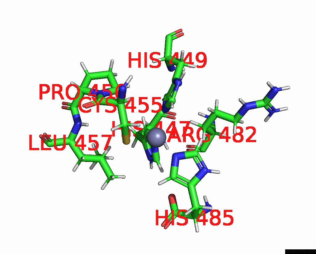
Mono view
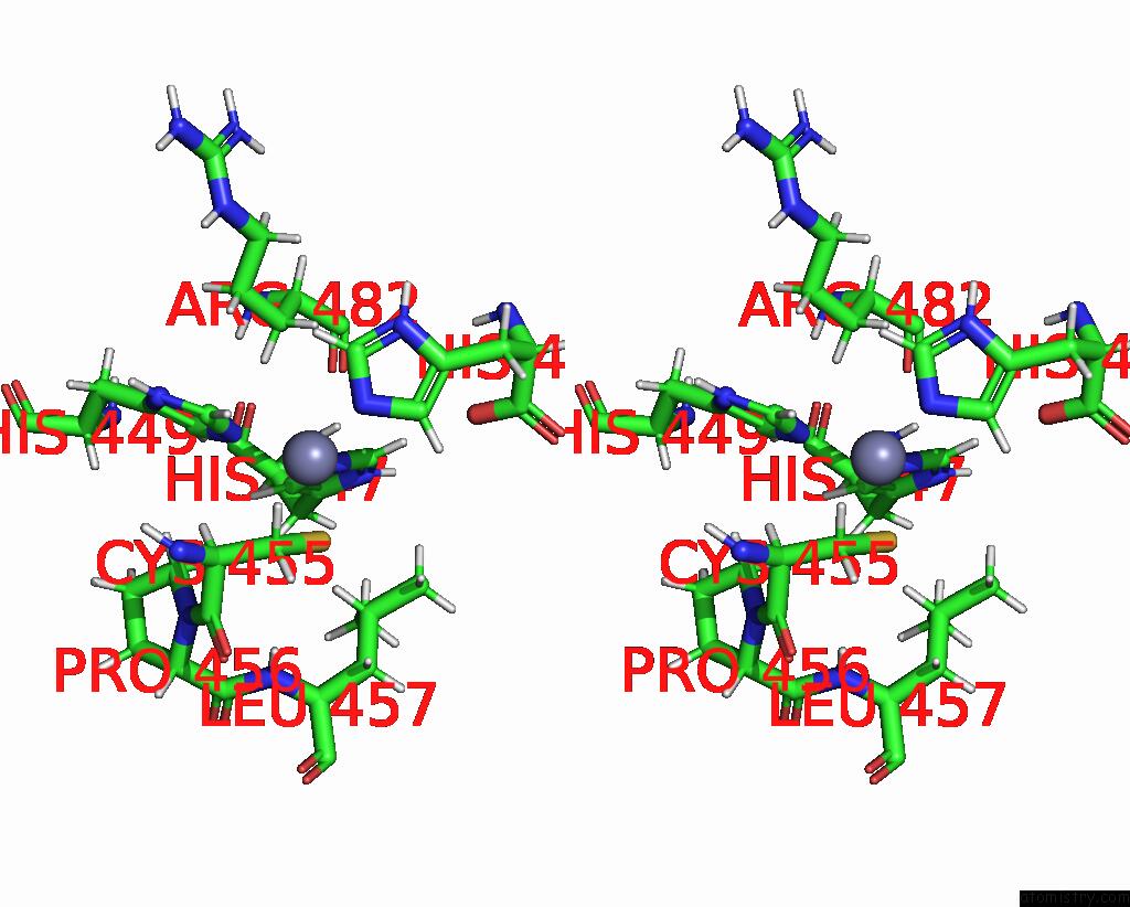
Stereo pair view
|
A full contact list of Zinc with other atoms in the Zn binding
site number 1 of Solution Structure of A Zinc-Binding Domain From the Junin Virus Envelope Glycoprotein within 5.0Å range:
probe
|
atom
|
residue
|
distance (Å)
|
B
|
Occ
|
A:Zn486
b:0.0
occ:1.00
|
NE2
|
A:HIS449
|
2.1
|
0.0
|
1.0
|
NE2
|
A:HIS485
|
2.1
|
0.0
|
1.0
|
NE2
|
A:HIS447
|
2.1
|
0.0
|
1.0
|
SG
|
A:CYS455
|
2.3
|
0.0
|
1.0
|
CE1
|
A:HIS485
|
3.1
|
0.0
|
1.0
|
CE1
|
A:HIS449
|
3.1
|
0.0
|
1.0
|
CE1
|
A:HIS447
|
3.1
|
0.0
|
1.0
|
CD2
|
A:HIS485
|
3.1
|
0.0
|
1.0
|
CD2
|
A:HIS449
|
3.1
|
0.0
|
1.0
|
CD2
|
A:HIS447
|
3.1
|
0.0
|
1.0
|
HB2
|
A:CYS455
|
3.2
|
0.0
|
1.0
|
HE1
|
A:HIS449
|
3.3
|
0.0
|
1.0
|
HE1
|
A:HIS485
|
3.3
|
0.0
|
1.0
|
HE1
|
A:HIS447
|
3.3
|
0.0
|
1.0
|
HD2
|
A:HIS485
|
3.3
|
0.0
|
1.0
|
CB
|
A:CYS455
|
3.3
|
0.0
|
1.0
|
HD2
|
A:HIS449
|
3.4
|
0.0
|
1.0
|
HD2
|
A:HIS447
|
3.4
|
0.0
|
1.0
|
HA
|
A:CYS455
|
3.4
|
0.0
|
1.0
|
HG
|
A:LEU457
|
3.7
|
0.0
|
1.0
|
HB2
|
A:ARG482
|
3.8
|
0.0
|
1.0
|
HB3
|
A:ARG482
|
3.9
|
0.0
|
1.0
|
HD2
|
A:PRO456
|
3.9
|
0.0
|
1.0
|
CA
|
A:CYS455
|
4.0
|
0.0
|
1.0
|
HD13
|
A:LEU457
|
4.1
|
0.0
|
1.0
|
HD3
|
A:PRO456
|
4.1
|
0.0
|
1.0
|
HD12
|
A:LEU457
|
4.2
|
0.0
|
1.0
|
ND1
|
A:HIS485
|
4.2
|
0.0
|
1.0
|
ND1
|
A:HIS449
|
4.2
|
0.0
|
1.0
|
CG
|
A:HIS485
|
4.2
|
0.0
|
1.0
|
ND1
|
A:HIS447
|
4.2
|
0.0
|
1.0
|
HB3
|
A:CYS455
|
4.3
|
0.0
|
1.0
|
CG
|
A:HIS449
|
4.3
|
0.0
|
1.0
|
CG
|
A:HIS447
|
4.3
|
0.0
|
1.0
|
CB
|
A:ARG482
|
4.3
|
0.0
|
1.0
|
HG3
|
A:ARG482
|
4.3
|
0.0
|
1.0
|
CD
|
A:PRO456
|
4.5
|
0.0
|
1.0
|
CD1
|
A:LEU457
|
4.5
|
0.0
|
1.0
|
CG
|
A:LEU457
|
4.6
|
0.0
|
1.0
|
OXT
|
A:HIS485
|
4.8
|
0.0
|
1.0
|
C
|
A:CYS455
|
4.8
|
0.0
|
1.0
|
CG
|
A:ARG482
|
4.9
|
0.0
|
1.0
|
H
|
A:LEU457
|
5.0
|
0.0
|
1.0
|
|
Zinc binding site 2 out
of 2 in 2l0z
Go back to
Zinc Binding Sites List in 2l0z
Zinc binding site 2 out
of 2 in the Solution Structure of A Zinc-Binding Domain From the Junin Virus Envelope Glycoprotein
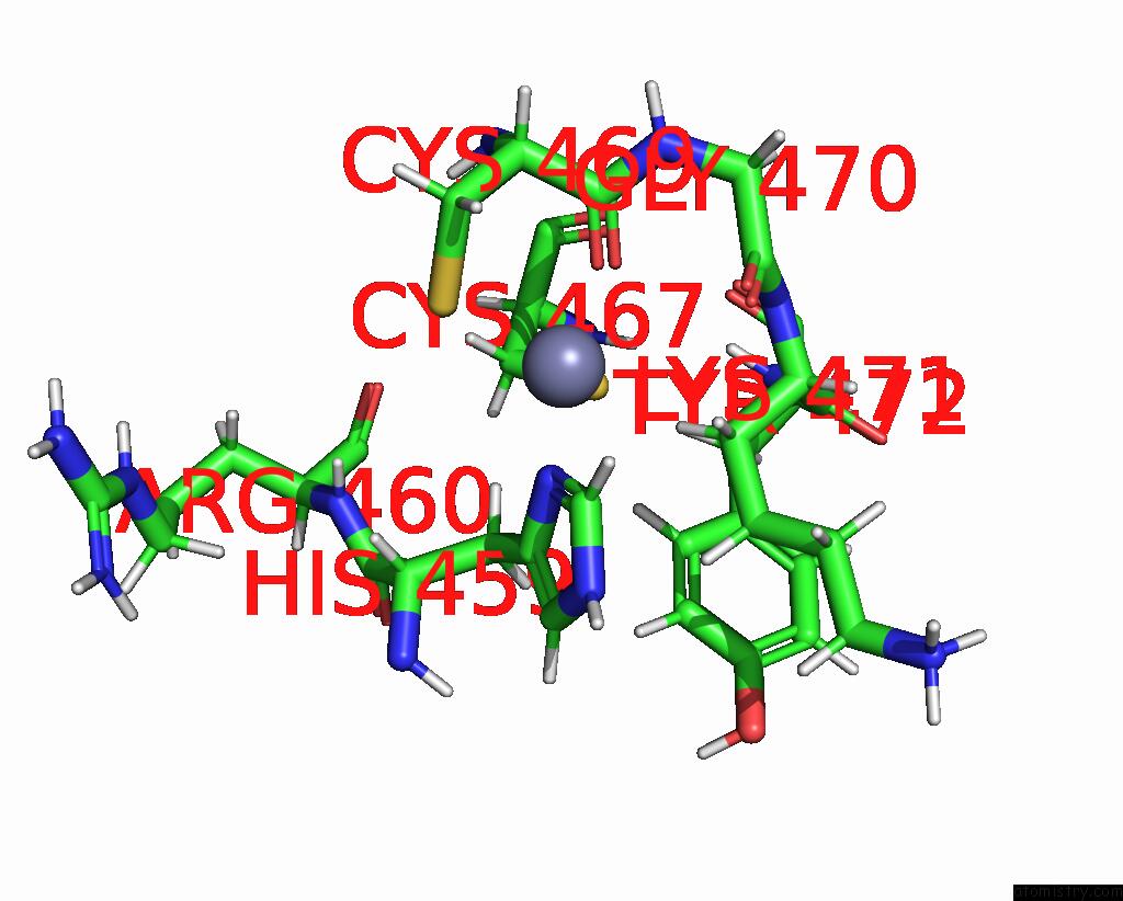
Mono view
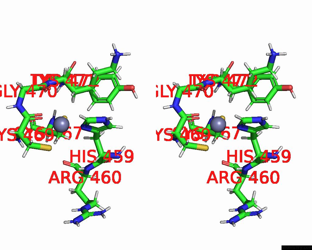
Stereo pair view
|
A full contact list of Zinc with other atoms in the Zn binding
site number 2 of Solution Structure of A Zinc-Binding Domain From the Junin Virus Envelope Glycoprotein within 5.0Å range:
probe
|
atom
|
residue
|
distance (Å)
|
B
|
Occ
|
A:Zn487
b:0.0
occ:1.00
|
O
|
A:CYS469
|
1.9
|
0.0
|
1.0
|
ND1
|
A:HIS459
|
2.1
|
0.0
|
1.0
|
SG
|
A:CYS469
|
2.3
|
0.0
|
1.0
|
SG
|
A:CYS467
|
2.3
|
0.0
|
1.0
|
HB3
|
A:LYS471
|
2.5
|
0.0
|
1.0
|
HB3
|
A:HIS459
|
2.9
|
0.0
|
1.0
|
HB2
|
A:LYS471
|
2.9
|
0.0
|
1.0
|
C
|
A:CYS469
|
3.0
|
0.0
|
1.0
|
CE1
|
A:HIS459
|
3.1
|
0.0
|
1.0
|
HB2
|
A:CYS467
|
3.1
|
0.0
|
1.0
|
CB
|
A:LYS471
|
3.1
|
0.0
|
1.0
|
CG
|
A:HIS459
|
3.1
|
0.0
|
1.0
|
HE1
|
A:HIS459
|
3.3
|
0.0
|
1.0
|
CB
|
A:CYS469
|
3.3
|
0.0
|
1.0
|
CB
|
A:CYS467
|
3.4
|
0.0
|
1.0
|
HB2
|
A:CYS469
|
3.4
|
0.0
|
1.0
|
CB
|
A:HIS459
|
3.5
|
0.0
|
1.0
|
O
|
A:GLY470
|
3.6
|
0.0
|
1.0
|
CA
|
A:CYS469
|
3.7
|
0.0
|
1.0
|
C
|
A:GLY470
|
3.7
|
0.0
|
1.0
|
N
|
A:LYS471
|
3.8
|
0.0
|
1.0
|
HB3
|
A:CYS467
|
4.0
|
0.0
|
1.0
|
HG2
|
A:LYS471
|
4.1
|
0.0
|
1.0
|
N
|
A:GLY470
|
4.1
|
0.0
|
1.0
|
CA
|
A:LYS471
|
4.1
|
0.0
|
1.0
|
H
|
A:TYR472
|
4.2
|
0.0
|
1.0
|
H
|
A:LYS471
|
4.2
|
0.0
|
1.0
|
HB2
|
A:HIS459
|
4.2
|
0.0
|
1.0
|
H
|
A:CYS469
|
4.2
|
0.0
|
1.0
|
NE2
|
A:HIS459
|
4.2
|
0.0
|
1.0
|
CG
|
A:LYS471
|
4.2
|
0.0
|
1.0
|
O
|
A:CYS467
|
4.2
|
0.0
|
1.0
|
N
|
A:CYS469
|
4.2
|
0.0
|
1.0
|
CD2
|
A:HIS459
|
4.3
|
0.0
|
1.0
|
HB3
|
A:CYS469
|
4.3
|
0.0
|
1.0
|
HA
|
A:HIS459
|
4.4
|
0.0
|
1.0
|
CA
|
A:GLY470
|
4.5
|
0.0
|
1.0
|
C
|
A:CYS467
|
4.5
|
0.0
|
1.0
|
CA
|
A:CYS467
|
4.6
|
0.0
|
1.0
|
HD2
|
A:TYR472
|
4.6
|
0.0
|
1.0
|
CA
|
A:HIS459
|
4.6
|
0.0
|
1.0
|
HA
|
A:CYS469
|
4.6
|
0.0
|
1.0
|
H
|
A:ARG460
|
4.7
|
0.0
|
1.0
|
HG3
|
A:LYS471
|
4.8
|
0.0
|
1.0
|
HA
|
A:LYS471
|
4.9
|
0.0
|
1.0
|
H
|
A:GLY470
|
4.9
|
0.0
|
1.0
|
N
|
A:TYR472
|
4.9
|
0.0
|
1.0
|
|
Reference:
K.Briknarova,
C.J.Thomas,
J.York,
J.H.Nunberg.
Structure of A Zinc-Binding Domain in the Junin Virus Envelope Glycoprotein. J.Biol.Chem. V. 286 1528 2011.
ISSN: ISSN 0021-9258
PubMed: 21068387
DOI: 10.1074/JBC.M110.166025
Page generated: Thu Oct 17 01:43:26 2024
|