Zinc »
PDB 2jb0-2jtn »
2jig »
Zinc in PDB 2jig: Crystal Structure of Chlamydomonas Reinhardtii Prolyl-4 Hydroxylase Type I Complexed with Zinc and Pyridine-2,4-Dicarboxylate
Protein crystallography data
The structure of Crystal Structure of Chlamydomonas Reinhardtii Prolyl-4 Hydroxylase Type I Complexed with Zinc and Pyridine-2,4-Dicarboxylate, PDB code: 2jig
was solved by
M.K.Koski,
R.Hieta,
C.Bollner,
K.I.Kivirikko,
J.Myllyharju,
R.K.Wierenga,
with X-Ray Crystallography technique. A brief refinement statistics is given in the table below:
| Resolution Low / High (Å) | 29.17 / 1.85 |
| Space group | P 21 21 21 |
| Cell size a, b, c (Å), α, β, γ (°) | 57.920, 60.250, 116.850, 90.00, 90.00, 90.00 |
| R / Rfree (%) | 18.3 / 23.8 |
Zinc Binding Sites:
The binding sites of Zinc atom in the Crystal Structure of Chlamydomonas Reinhardtii Prolyl-4 Hydroxylase Type I Complexed with Zinc and Pyridine-2,4-Dicarboxylate
(pdb code 2jig). This binding sites where shown within
5.0 Angstroms radius around Zinc atom.
In total 4 binding sites of Zinc where determined in the Crystal Structure of Chlamydomonas Reinhardtii Prolyl-4 Hydroxylase Type I Complexed with Zinc and Pyridine-2,4-Dicarboxylate, PDB code: 2jig:
Jump to Zinc binding site number: 1; 2; 3; 4;
In total 4 binding sites of Zinc where determined in the Crystal Structure of Chlamydomonas Reinhardtii Prolyl-4 Hydroxylase Type I Complexed with Zinc and Pyridine-2,4-Dicarboxylate, PDB code: 2jig:
Jump to Zinc binding site number: 1; 2; 3; 4;
Zinc binding site 1 out of 4 in 2jig
Go back to
Zinc binding site 1 out
of 4 in the Crystal Structure of Chlamydomonas Reinhardtii Prolyl-4 Hydroxylase Type I Complexed with Zinc and Pyridine-2,4-Dicarboxylate
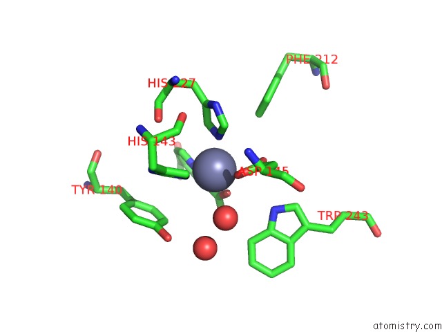
Mono view

Stereo pair view
Mono view
Stereo pair view
A full contact list of Zinc with other atoms in the Zn binding
site number 1 of Crystal Structure of Chlamydomonas Reinhardtii Prolyl-4 Hydroxylase Type I Complexed with Zinc and Pyridine-2,4-Dicarboxylate within 5.0Å range:
|
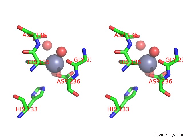
Zinc binding site 2 out of 4 in 2jig
Go back to
Zinc binding site 2 out
of 4 in the Crystal Structure of Chlamydomonas Reinhardtii Prolyl-4 Hydroxylase Type I Complexed with Zinc and Pyridine-2,4-Dicarboxylate

Mono view
Stereo pair view
Mono view
Stereo pair view
A full contact list of Zinc with other atoms in the Zn binding
site number 2 of Crystal Structure of Chlamydomonas Reinhardtii Prolyl-4 Hydroxylase Type I Complexed with Zinc and Pyridine-2,4-Dicarboxylate within 5.0Å range:
|
Zinc binding site 3 out of 4 in 2jig
Go back to
Zinc binding site 3 out
of 4 in the Crystal Structure of Chlamydomonas Reinhardtii Prolyl-4 Hydroxylase Type I Complexed with Zinc and Pyridine-2,4-Dicarboxylate
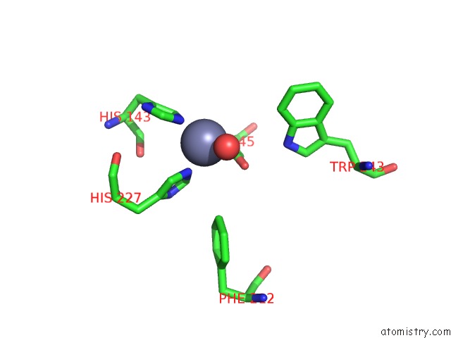
Mono view

Stereo pair view
Mono view
Stereo pair view
A full contact list of Zinc with other atoms in the Zn binding
site number 3 of Crystal Structure of Chlamydomonas Reinhardtii Prolyl-4 Hydroxylase Type I Complexed with Zinc and Pyridine-2,4-Dicarboxylate within 5.0Å range:
|
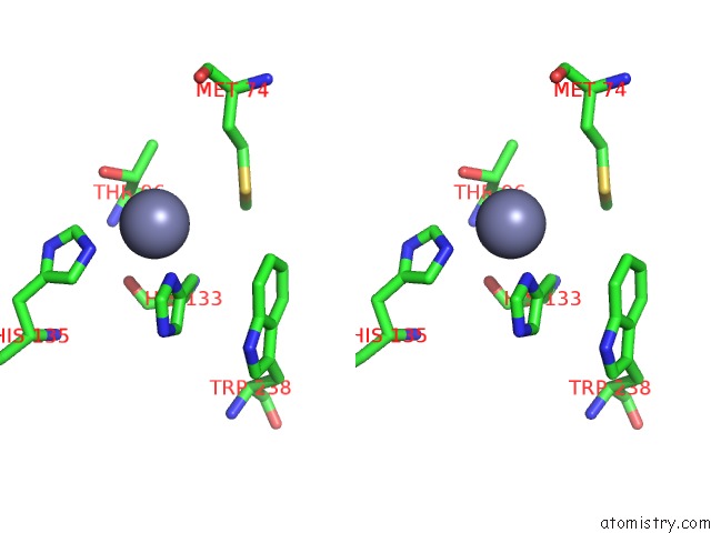
Zinc binding site 4 out of 4 in 2jig
Go back to
Zinc binding site 4 out
of 4 in the Crystal Structure of Chlamydomonas Reinhardtii Prolyl-4 Hydroxylase Type I Complexed with Zinc and Pyridine-2,4-Dicarboxylate

Mono view
Stereo pair view
Mono view
Stereo pair view
A full contact list of Zinc with other atoms in the Zn binding
site number 4 of Crystal Structure of Chlamydomonas Reinhardtii Prolyl-4 Hydroxylase Type I Complexed with Zinc and Pyridine-2,4-Dicarboxylate within 5.0Å range:
|
Reference:
M.K.Koski,
R.Hieta,
C.Bollner,
K.I.Kivirikko,
J.Myllyharju,
R.K.Wierenga.
The Active Site of An Algal Prolyl 4-Hydroxylase Has A Large Structural Plasticity. J.Biol.Chem. V. 282 37112 2007.
ISSN: ISSN 0021-9258
PubMed: 17940281
DOI: 10.1074/JBC.M706554200
Page generated: Wed Aug 20 03:48:16 2025
ISSN: ISSN 0021-9258
PubMed: 17940281
DOI: 10.1074/JBC.M706554200
Last articles
Zn in 3D7SZn in 3D7G
Zn in 3D7H
Zn in 3D7D
Zn in 3D7F
Zn in 3D68
Zn in 3D67
Zn in 3D66
Zn in 3D71
Zn in 3D6N