Zinc »
PDB 2jb0-2jtn »
2jhg »
Zinc in PDB 2jhg: Structural Evidence For A Ligand Coordination Switch in Liver Alcohol Dehydrogenase
Enzymatic activity of Structural Evidence For A Ligand Coordination Switch in Liver Alcohol Dehydrogenase
All present enzymatic activity of Structural Evidence For A Ligand Coordination Switch in Liver Alcohol Dehydrogenase:
1.1.1.1;
1.1.1.1;
Protein crystallography data
The structure of Structural Evidence For A Ligand Coordination Switch in Liver Alcohol Dehydrogenase, PDB code: 2jhg
was solved by
R.Meijers,
H.W.Adolph,
Z.Dauter,
K.S.Wilson,
V.S.Lamzin,
E.S.Cedergren-Zeppezauer,
with X-Ray Crystallography technique. A brief refinement statistics is given in the table below:
| Resolution Low / High (Å) | 19.83 / 1.20 |
| Space group | P 1 |
| Cell size a, b, c (Å), α, β, γ (°) | 50.186, 43.798, 92.516, 77.12, 87.44, 108.89 |
| R / Rfree (%) | 11.4 / 14.2 |
Zinc Binding Sites:
The binding sites of Zinc atom in the Structural Evidence For A Ligand Coordination Switch in Liver Alcohol Dehydrogenase
(pdb code 2jhg). This binding sites where shown within
5.0 Angstroms radius around Zinc atom.
In total 4 binding sites of Zinc where determined in the Structural Evidence For A Ligand Coordination Switch in Liver Alcohol Dehydrogenase, PDB code: 2jhg:
Jump to Zinc binding site number: 1; 2; 3; 4;
In total 4 binding sites of Zinc where determined in the Structural Evidence For A Ligand Coordination Switch in Liver Alcohol Dehydrogenase, PDB code: 2jhg:
Jump to Zinc binding site number: 1; 2; 3; 4;
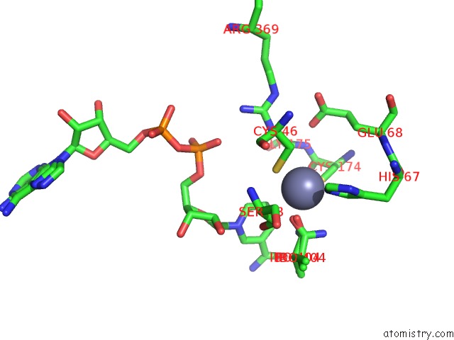
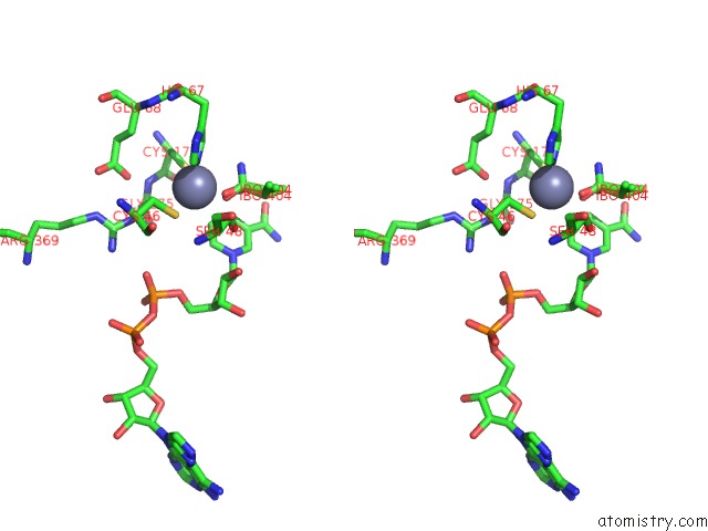
Zinc binding site 1 out of 4 in 2jhg
Go back to
Zinc binding site 1 out
of 4 in the Structural Evidence For A Ligand Coordination Switch in Liver Alcohol Dehydrogenase

Mono view
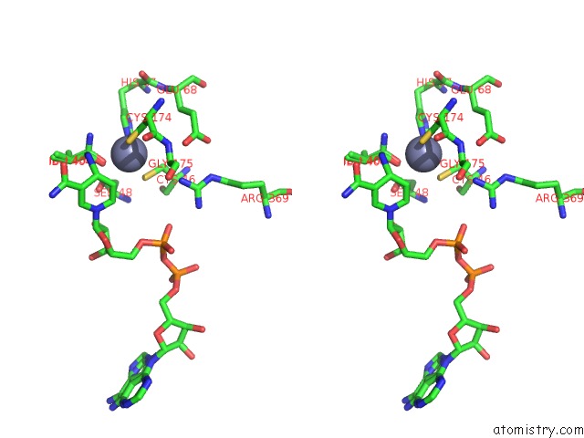
Stereo pair view
Mono view
Stereo pair view
A full contact list of Zinc with other atoms in the Zn binding
site number 1 of Structural Evidence For A Ligand Coordination Switch in Liver Alcohol Dehydrogenase within 5.0Å range:
|
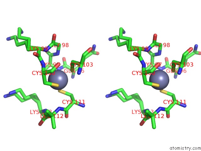
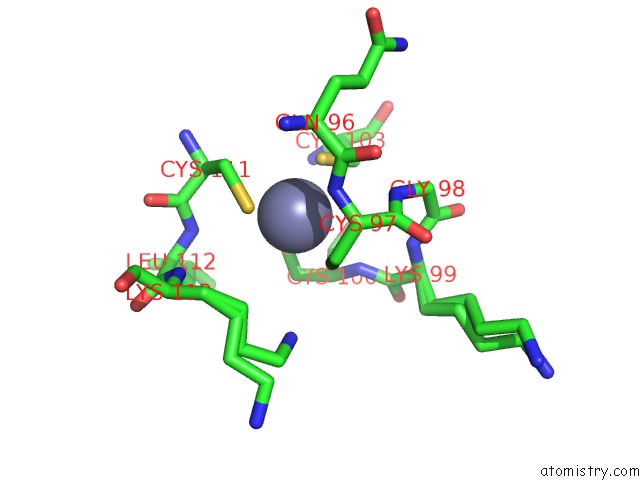
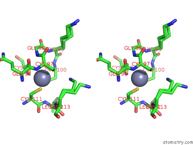
Zinc binding site 2 out of 4 in 2jhg
Go back to
Zinc binding site 2 out
of 4 in the Structural Evidence For A Ligand Coordination Switch in Liver Alcohol Dehydrogenase
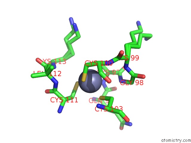
Mono view
Stereo pair view
Mono view
Stereo pair view
A full contact list of Zinc with other atoms in the Zn binding
site number 2 of Structural Evidence For A Ligand Coordination Switch in Liver Alcohol Dehydrogenase within 5.0Å range:
|
Zinc binding site 3 out of 4 in 2jhg
Go back to
Zinc binding site 3 out
of 4 in the Structural Evidence For A Ligand Coordination Switch in Liver Alcohol Dehydrogenase
Mono view
Stereo pair view
Mono view
Stereo pair view
A full contact list of Zinc with other atoms in the Zn binding
site number 3 of Structural Evidence For A Ligand Coordination Switch in Liver Alcohol Dehydrogenase within 5.0Å range:
|
Zinc binding site 4 out of 4 in 2jhg
Go back to
Zinc binding site 4 out
of 4 in the Structural Evidence For A Ligand Coordination Switch in Liver Alcohol Dehydrogenase
Mono view
Stereo pair view
Mono view
Stereo pair view
A full contact list of Zinc with other atoms in the Zn binding
site number 4 of Structural Evidence For A Ligand Coordination Switch in Liver Alcohol Dehydrogenase within 5.0Å range:
|
Reference:
R.Meijers,
H.W.Adolph,
Z.Dauter,
K.S.Wilson,
V.S.Lamzin,
E.S.Cedergren-Zeppezauer.
Structural Evidence For A Ligand Coordination Switch in Liver Alcohol Dehydrogenase Biochemistry V. 46 5446 2007.
ISSN: ISSN 0006-2960
PubMed: 17429946
DOI: 10.1021/BI6023594
Page generated: Thu Oct 17 01:13:47 2024
ISSN: ISSN 0006-2960
PubMed: 17429946
DOI: 10.1021/BI6023594
Last articles
Ag in 7XLVAg in 8DX7
Ag in 8DX6
Ag in 7XKM
Ag in 8DX5
Ag in 8DX1
Ag in 8C2Q
Ag in 7WAA
Ag in 7TMJ
Ag in 7TGD