Zinc »
PDB 2ivh-2jaz »
2ivh »
Zinc in PDB 2ivh: Crystal Structure of the Nuclease Domain of COLE7 (H545Q Mutant) in Complex with An 18-Bp Duplex Dna
Protein crystallography data
The structure of Crystal Structure of the Nuclease Domain of COLE7 (H545Q Mutant) in Complex with An 18-Bp Duplex Dna, PDB code: 2ivh
was solved by
Y.T.Wang,
W.J.Yang,
C.L.Li,
L.G.Doudeva,
H.S.Yuan,
with X-Ray Crystallography technique. A brief refinement statistics is given in the table below:
| Resolution Low / High (Å) | 47.76 / 2.80 |
| Space group | P 41 21 2 |
| Cell size a, b, c (Å), α, β, γ (°) | 106.800, 106.800, 60.200, 90.00, 90.00, 90.00 |
| R / Rfree (%) | 19.3 / 26.5 |
Zinc Binding Sites:
The binding sites of Zinc atom in the Crystal Structure of the Nuclease Domain of COLE7 (H545Q Mutant) in Complex with An 18-Bp Duplex Dna
(pdb code 2ivh). This binding sites where shown within
5.0 Angstroms radius around Zinc atom.
In total only one binding site of Zinc was determined in the Crystal Structure of the Nuclease Domain of COLE7 (H545Q Mutant) in Complex with An 18-Bp Duplex Dna, PDB code: 2ivh:
In total only one binding site of Zinc was determined in the Crystal Structure of the Nuclease Domain of COLE7 (H545Q Mutant) in Complex with An 18-Bp Duplex Dna, PDB code: 2ivh:
Zinc binding site 1 out of 1 in 2ivh
Go back to
Zinc binding site 1 out
of 1 in the Crystal Structure of the Nuclease Domain of COLE7 (H545Q Mutant) in Complex with An 18-Bp Duplex Dna
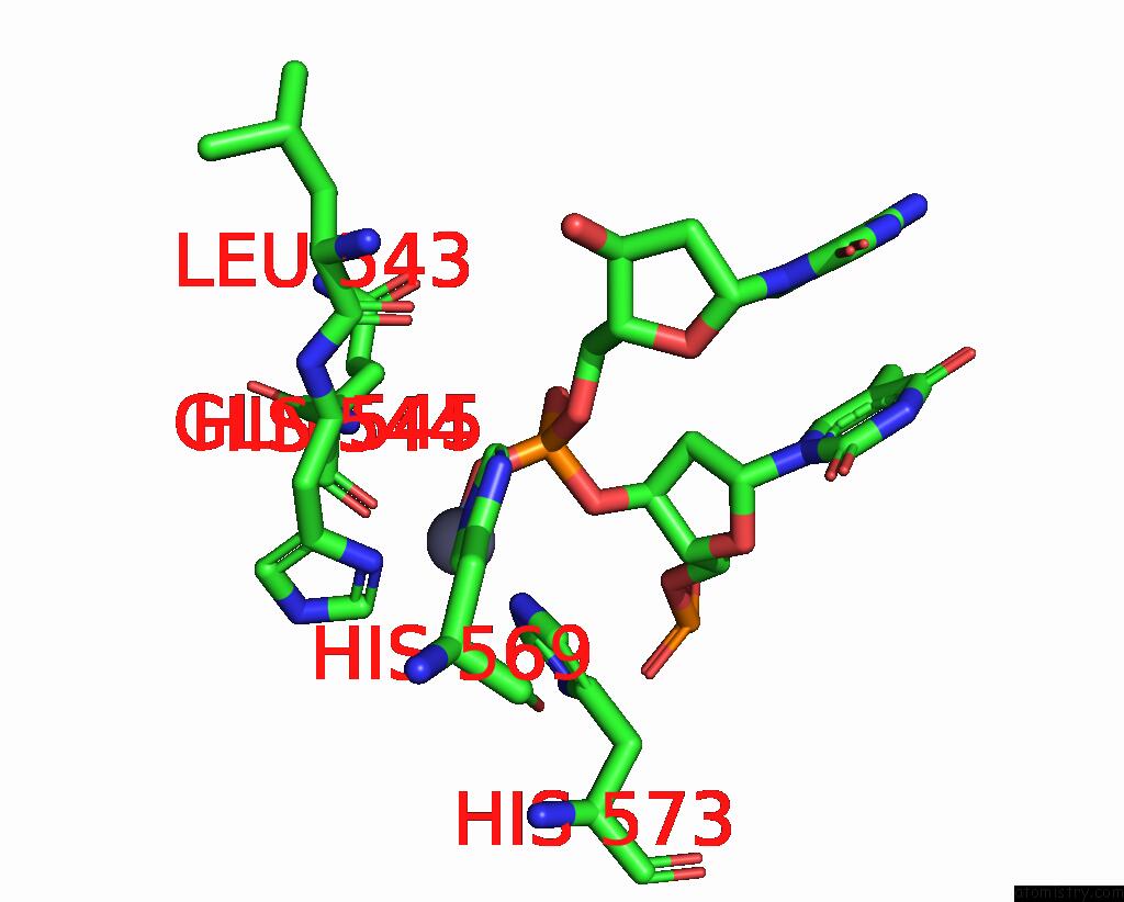
Mono view
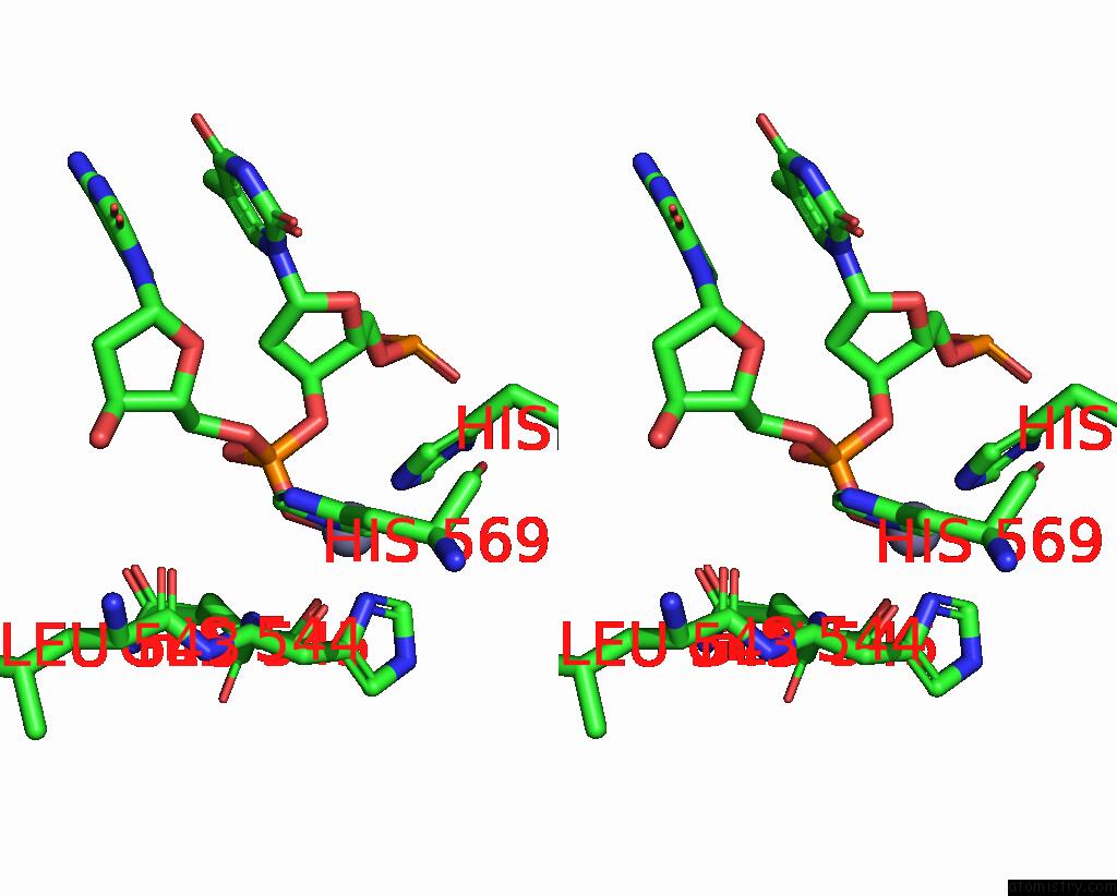
Stereo pair view
Mono view
Stereo pair view
A full contact list of Zinc with other atoms in the Zn binding
site number 1 of Crystal Structure of the Nuclease Domain of COLE7 (H545Q Mutant) in Complex with An 18-Bp Duplex Dna within 5.0Å range:
|
Reference:
Y.T.Wang,
W.J.Yang,
C.L.Li,
L.G.Doudeva,
H.S.Yuan.
Structural Basis For Sequence-Dependent Dna Cleavage By Nonspecific Endonucleases. Nucleic Acids Res. V. 35 584 2007.
ISSN: ISSN 0305-1048
PubMed: 17175542
DOI: 10.1093/NAR/GKL621
Page generated: Thu Oct 17 01:00:29 2024
ISSN: ISSN 0305-1048
PubMed: 17175542
DOI: 10.1093/NAR/GKL621
Last articles
Al in 6S3MAl in 6S9E
Al in 6S3H
Al in 6NJP
Al in 6NJO
Al in 6QP0
Al in 6L3G
Al in 6K7M
Al in 6K7N
Al in 6JIM