Zinc »
PDB 2ics-2iv0 »
2iq6 »
Zinc in PDB 2iq6: Crystal Structure of the Aminopeptidase From Vibrio Proteolyticus in Complexation with Leucyl-Leucyl-Leucine.
Enzymatic activity of Crystal Structure of the Aminopeptidase From Vibrio Proteolyticus in Complexation with Leucyl-Leucyl-Leucine.
All present enzymatic activity of Crystal Structure of the Aminopeptidase From Vibrio Proteolyticus in Complexation with Leucyl-Leucyl-Leucine.:
3.4.11.10;
3.4.11.10;
Protein crystallography data
The structure of Crystal Structure of the Aminopeptidase From Vibrio Proteolyticus in Complexation with Leucyl-Leucyl-Leucine., PDB code: 2iq6
was solved by
A.Kumar,
B.Narayanan,
J.-J.P.Kim,
B.Bennett,
with X-Ray Crystallography technique. A brief refinement statistics is given in the table below:
| Resolution Low / High (Å) | 30.00 / 2.00 |
| Space group | P 61 2 2 |
| Cell size a, b, c (Å), α, β, γ (°) | 108.400, 108.400, 96.800, 90.00, 90.00, 120.00 |
| R / Rfree (%) | 20.6 / 24.7 |
Zinc Binding Sites:
The binding sites of Zinc atom in the Crystal Structure of the Aminopeptidase From Vibrio Proteolyticus in Complexation with Leucyl-Leucyl-Leucine.
(pdb code 2iq6). This binding sites where shown within
5.0 Angstroms radius around Zinc atom.
In total 2 binding sites of Zinc where determined in the Crystal Structure of the Aminopeptidase From Vibrio Proteolyticus in Complexation with Leucyl-Leucyl-Leucine., PDB code: 2iq6:
Jump to Zinc binding site number: 1; 2;
In total 2 binding sites of Zinc where determined in the Crystal Structure of the Aminopeptidase From Vibrio Proteolyticus in Complexation with Leucyl-Leucyl-Leucine., PDB code: 2iq6:
Jump to Zinc binding site number: 1; 2;
Zinc binding site 1 out of 2 in 2iq6
Go back to
Zinc binding site 1 out
of 2 in the Crystal Structure of the Aminopeptidase From Vibrio Proteolyticus in Complexation with Leucyl-Leucyl-Leucine.
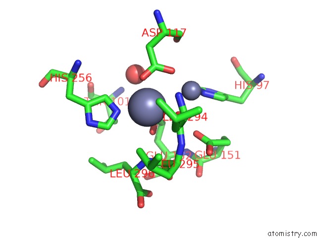
Mono view
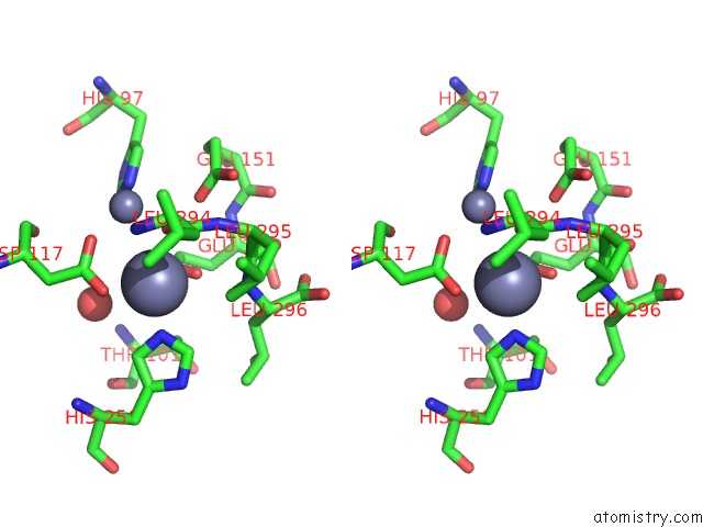
Stereo pair view
Mono view
Stereo pair view
A full contact list of Zinc with other atoms in the Zn binding
site number 1 of Crystal Structure of the Aminopeptidase From Vibrio Proteolyticus in Complexation with Leucyl-Leucyl-Leucine. within 5.0Å range:
|
Zinc binding site 2 out of 2 in 2iq6
Go back to
Zinc binding site 2 out
of 2 in the Crystal Structure of the Aminopeptidase From Vibrio Proteolyticus in Complexation with Leucyl-Leucyl-Leucine.
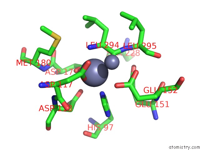
Mono view
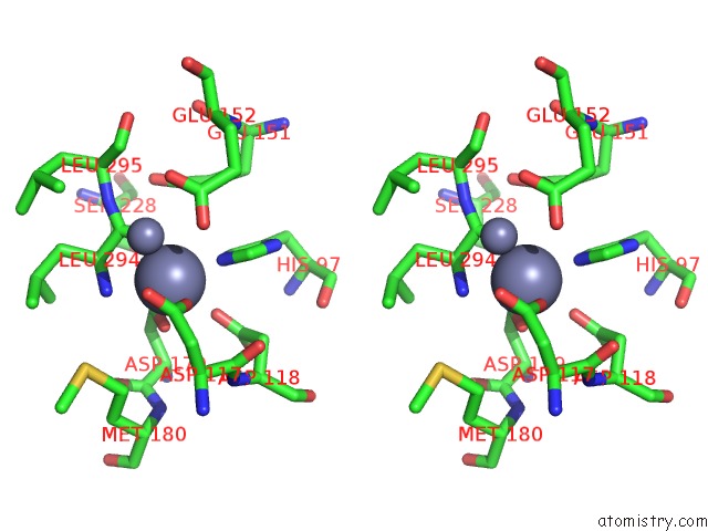
Stereo pair view
Mono view
Stereo pair view
A full contact list of Zinc with other atoms in the Zn binding
site number 2 of Crystal Structure of the Aminopeptidase From Vibrio Proteolyticus in Complexation with Leucyl-Leucyl-Leucine. within 5.0Å range:
|
Reference:
A.Kumar,
G.R.Periyannan,
B.Narayanan,
A.W.Kittell,
J.-J.Kim,
B.Bennett.
Experimental Evidence For A Metallohydrolase Mechanism in Which the Nucleophile Is Not Delivered By A Metal Ion: Epr Spectrokinetic and Structural Studies of Aminopeptidase From Vibrio Proteolyticus Biochem.J. V. 403 527 2007.
ISSN: ISSN 0264-6021
PubMed: 17238863
DOI: 10.1042/BJ20061591
Page generated: Thu Oct 17 00:58:10 2024
ISSN: ISSN 0264-6021
PubMed: 17238863
DOI: 10.1042/BJ20061591
Last articles
Zn in 9MJ5Zn in 9HNW
Zn in 9G0L
Zn in 9FNE
Zn in 9DZN
Zn in 9E0I
Zn in 9D32
Zn in 9DAK
Zn in 8ZXC
Zn in 8ZUF