Zinc »
PDB 2ics-2iv0 »
2ioi »
Zinc in PDB 2ioi: Crystal Structure of the Mouse P53 Core Domain at 1.55 A
Protein crystallography data
The structure of Crystal Structure of the Mouse P53 Core Domain at 1.55 A, PDB code: 2ioi
was solved by
W.C.Ho,
C.Luo,
K.Zhao,
X.Chai,
M.X.Fitzgerald,
R.Marmorstein,
with X-Ray Crystallography technique. A brief refinement statistics is given in the table below:
| Resolution Low / High (Å) | 30.00 / 1.55 |
| Space group | C 1 2 1 |
| Cell size a, b, c (Å), α, β, γ (°) | 92.316, 44.688, 63.051, 90.00, 126.25, 90.00 |
| R / Rfree (%) | 18.4 / 22.8 |
Zinc Binding Sites:
The binding sites of Zinc atom in the Crystal Structure of the Mouse P53 Core Domain at 1.55 A
(pdb code 2ioi). This binding sites where shown within
5.0 Angstroms radius around Zinc atom.
In total only one binding site of Zinc was determined in the Crystal Structure of the Mouse P53 Core Domain at 1.55 A, PDB code: 2ioi:
In total only one binding site of Zinc was determined in the Crystal Structure of the Mouse P53 Core Domain at 1.55 A, PDB code: 2ioi:
Zinc binding site 1 out of 1 in 2ioi
Go back to
Zinc binding site 1 out
of 1 in the Crystal Structure of the Mouse P53 Core Domain at 1.55 A
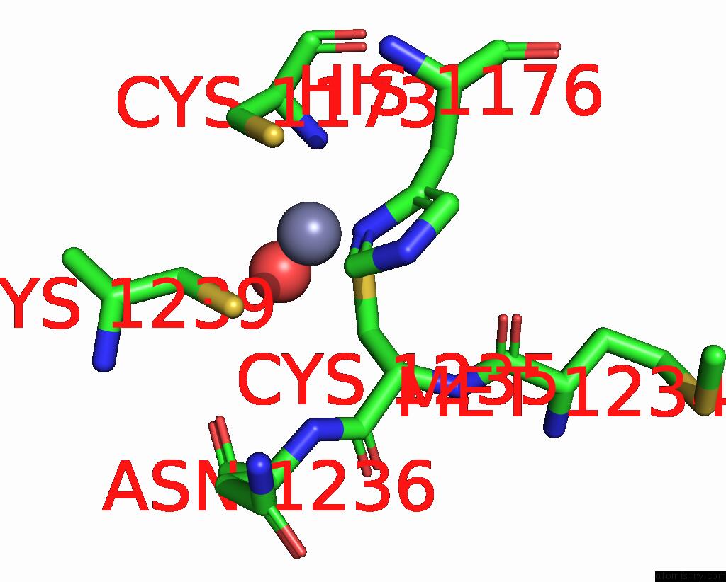
Mono view
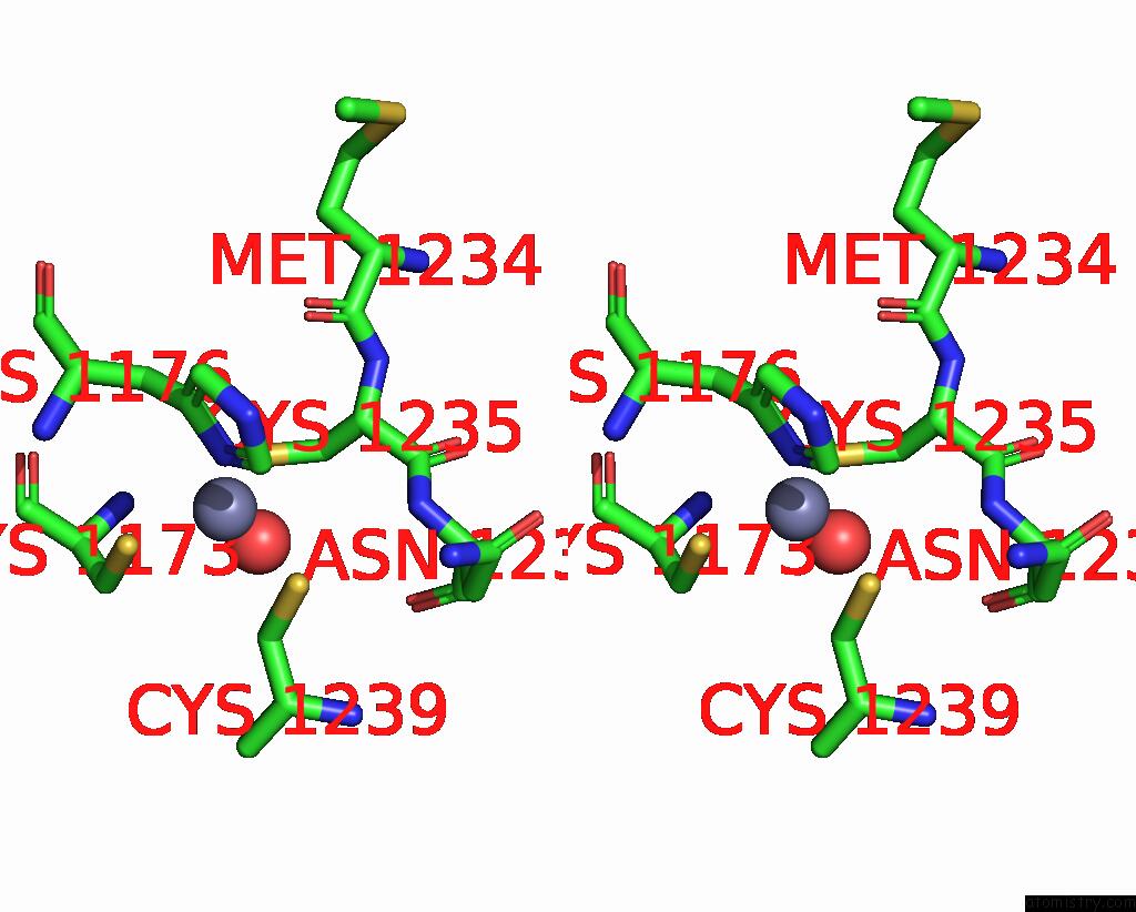
Stereo pair view
Mono view
Stereo pair view
A full contact list of Zinc with other atoms in the Zn binding
site number 1 of Crystal Structure of the Mouse P53 Core Domain at 1.55 A within 5.0Å range:
|
Reference:
W.C.Ho,
C.Luo,
K.Zhao,
X.Chai,
M.X.Fitzgerald,
R.Marmorstein.
High-Resolution Structure of the P53 Core Domain: Implications For Binding Small-Molecule Stabilizing Compounds. Acta Crystallogr.,Sect.D V. 62 1484 2006.
ISSN: ISSN 0907-4449
PubMed: 17139084
DOI: 10.1107/S090744490603890X
Page generated: Thu Oct 17 00:57:50 2024
ISSN: ISSN 0907-4449
PubMed: 17139084
DOI: 10.1107/S090744490603890X
Last articles
Zn in 9MJ5Zn in 9HNW
Zn in 9G0L
Zn in 9FNE
Zn in 9DZN
Zn in 9E0I
Zn in 9D32
Zn in 9DAK
Zn in 8ZXC
Zn in 8ZUF