Zinc »
PDB 2ics-2iv0 »
2inn »
Zinc in PDB 2inn: Structure of the Phenol Hydroxyalse-Regulatory Protein Complex
Protein crystallography data
The structure of Structure of the Phenol Hydroxyalse-Regulatory Protein Complex, PDB code: 2inn
was solved by
M.H.Sazinsky,
P.W.Dunten,
M.S.Mccormick,
S.J.Lippard,
with X-Ray Crystallography technique. A brief refinement statistics is given in the table below:
| Resolution Low / High (Å) | 30.00 / 2.70 |
| Space group | P 21 21 21 |
| Cell size a, b, c (Å), α, β, γ (°) | 87.453, 146.925, 189.787, 90.00, 90.00, 90.00 |
| R / Rfree (%) | 20.2 / 25.2 |
Other elements in 2inn:
The structure of Structure of the Phenol Hydroxyalse-Regulatory Protein Complex also contains other interesting chemical elements:
| Molybdenum | (Mo) | 1 atom |
| Iron | (Fe) | 4 atoms |
Zinc Binding Sites:
The binding sites of Zinc atom in the Structure of the Phenol Hydroxyalse-Regulatory Protein Complex
(pdb code 2inn). This binding sites where shown within
5.0 Angstroms radius around Zinc atom.
In total 2 binding sites of Zinc where determined in the Structure of the Phenol Hydroxyalse-Regulatory Protein Complex, PDB code: 2inn:
Jump to Zinc binding site number: 1; 2;
In total 2 binding sites of Zinc where determined in the Structure of the Phenol Hydroxyalse-Regulatory Protein Complex, PDB code: 2inn:
Jump to Zinc binding site number: 1; 2;
Zinc binding site 1 out of 2 in 2inn
Go back to
Zinc binding site 1 out
of 2 in the Structure of the Phenol Hydroxyalse-Regulatory Protein Complex
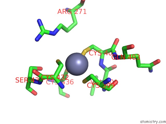
Mono view
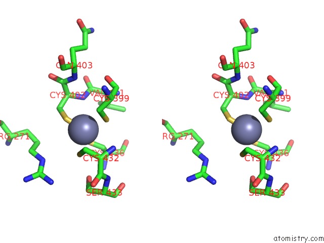
Stereo pair view
Mono view
Stereo pair view
A full contact list of Zinc with other atoms in the Zn binding
site number 1 of Structure of the Phenol Hydroxyalse-Regulatory Protein Complex within 5.0Å range:
|
Zinc binding site 2 out of 2 in 2inn
Go back to
Zinc binding site 2 out
of 2 in the Structure of the Phenol Hydroxyalse-Regulatory Protein Complex
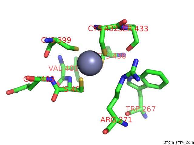
Mono view
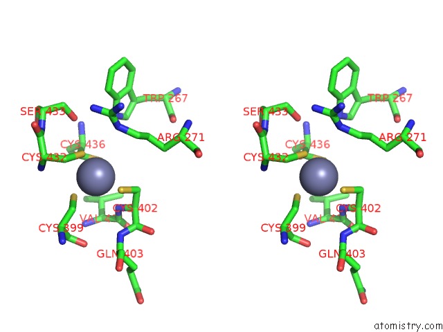
Stereo pair view
Mono view
Stereo pair view
A full contact list of Zinc with other atoms in the Zn binding
site number 2 of Structure of the Phenol Hydroxyalse-Regulatory Protein Complex within 5.0Å range:
|
Reference:
M.H.Sazinsky,
P.W.Dunten,
M.S.Mccormick,
A.Didonato,
S.J.Lippard.
X-Ray Structure of A Hydroxylase-Regulatory Protein Complex From A Hydrocarbon-Oxidizing Multicomponent Monooxygenase, Pseudomonas Sp. OX1 Phenol Hydroxylase. Biochemistry V. 45 15392 2006.
ISSN: ISSN 0006-2960
PubMed: 17176061
DOI: 10.1021/BI0618969
Page generated: Thu Oct 17 00:57:00 2024
ISSN: ISSN 0006-2960
PubMed: 17176061
DOI: 10.1021/BI0618969
Last articles
Zn in 9MJ5Zn in 9HNW
Zn in 9G0L
Zn in 9FNE
Zn in 9DZN
Zn in 9E0I
Zn in 9D32
Zn in 9DAK
Zn in 8ZXC
Zn in 8ZUF