Zinc »
PDB 2ics-2iv0 »
2ili »
Zinc in PDB 2ili: Refine Atomic Structure of Human Carbonic Anhydrase II
Enzymatic activity of Refine Atomic Structure of Human Carbonic Anhydrase II
All present enzymatic activity of Refine Atomic Structure of Human Carbonic Anhydrase II:
4.2.1.1;
4.2.1.1;
Protein crystallography data
The structure of Refine Atomic Structure of Human Carbonic Anhydrase II, PDB code: 2ili
was solved by
S.Z.Fisher,
with X-Ray Crystallography technique. A brief refinement statistics is given in the table below:
| Resolution Low / High (Å) | 20.00 / 1.05 |
| Space group | P 1 21 1 |
| Cell size a, b, c (Å), α, β, γ (°) | 42.212, 41.291, 72.154, 90.00, 104.39, 90.00 |
| R / Rfree (%) | 12 / 15.1 |
Zinc Binding Sites:
The binding sites of Zinc atom in the Refine Atomic Structure of Human Carbonic Anhydrase II
(pdb code 2ili). This binding sites where shown within
5.0 Angstroms radius around Zinc atom.
In total only one binding site of Zinc was determined in the Refine Atomic Structure of Human Carbonic Anhydrase II, PDB code: 2ili:
In total only one binding site of Zinc was determined in the Refine Atomic Structure of Human Carbonic Anhydrase II, PDB code: 2ili:
Zinc binding site 1 out of 1 in 2ili
Go back to
Zinc binding site 1 out
of 1 in the Refine Atomic Structure of Human Carbonic Anhydrase II
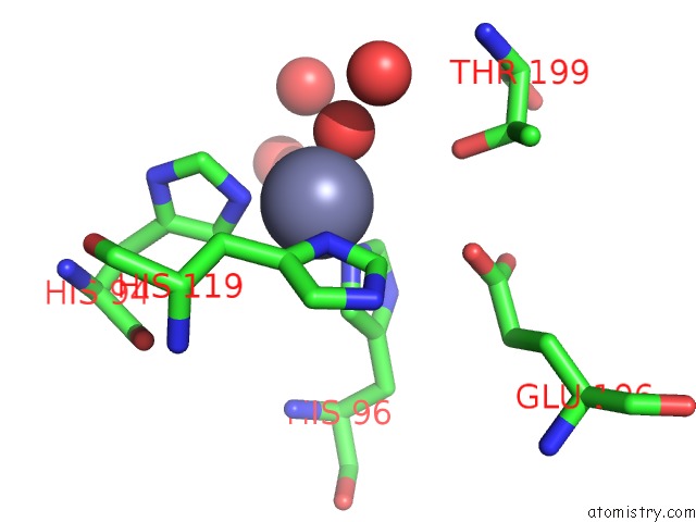
Mono view
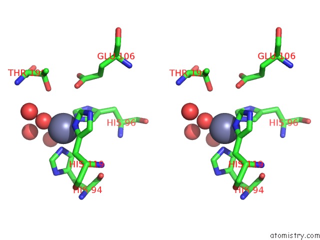
Stereo pair view
Mono view
Stereo pair view
A full contact list of Zinc with other atoms in the Zn binding
site number 1 of Refine Atomic Structure of Human Carbonic Anhydrase II within 5.0Å range:
|
Reference:
S.Z.Fisher,
C.M.Maupin,
M.Budayova-Spano,
L.Govindasamy,
C.Tu,
M.Agbandje-Mckenna,
D.N.Silverman,
G.A.Voth,
R.Mckenna.
Atomic Crystal and Molecular Dynamics Simulation Structures of Human Carbonic Anhydrase II: Insights Into the Proton Transfer Mechanism Biochemistry V. 46 2930 2007.
ISSN: ISSN 0006-2960
PubMed: 17319692
DOI: 10.1021/BI062066Y
Page generated: Thu Oct 17 00:55:31 2024
ISSN: ISSN 0006-2960
PubMed: 17319692
DOI: 10.1021/BI062066Y
Last articles
Zn in 9MJ5Zn in 9HNW
Zn in 9G0L
Zn in 9FNE
Zn in 9DZN
Zn in 9E0I
Zn in 9D32
Zn in 9DAK
Zn in 8ZXC
Zn in 8ZUF