Zinc »
PDB 2hqk-2ibi »
2i3i »
Zinc in PDB 2i3i: Structure of An Ml-Iap/Xiap Chimera Bound to A Peptidomimetic
Protein crystallography data
The structure of Structure of An Ml-Iap/Xiap Chimera Bound to A Peptidomimetic, PDB code: 2i3i
was solved by
W.J.Fairbrother,
M.C.Franklin,
with X-Ray Crystallography technique. A brief refinement statistics is given in the table below:
| Resolution Low / High (Å) | 24.34 / 2.30 |
| Space group | P 41 21 2 |
| Cell size a, b, c (Å), α, β, γ (°) | 87.356, 87.356, 73.196, 90.00, 90.00, 90.00 |
| R / Rfree (%) | 18.8 / 22.4 |
Zinc Binding Sites:
The binding sites of Zinc atom in the Structure of An Ml-Iap/Xiap Chimera Bound to A Peptidomimetic
(pdb code 2i3i). This binding sites where shown within
5.0 Angstroms radius around Zinc atom.
In total 2 binding sites of Zinc where determined in the Structure of An Ml-Iap/Xiap Chimera Bound to A Peptidomimetic, PDB code: 2i3i:
Jump to Zinc binding site number: 1; 2;
In total 2 binding sites of Zinc where determined in the Structure of An Ml-Iap/Xiap Chimera Bound to A Peptidomimetic, PDB code: 2i3i:
Jump to Zinc binding site number: 1; 2;
Zinc binding site 1 out of 2 in 2i3i
Go back to
Zinc binding site 1 out
of 2 in the Structure of An Ml-Iap/Xiap Chimera Bound to A Peptidomimetic
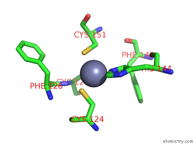
Mono view
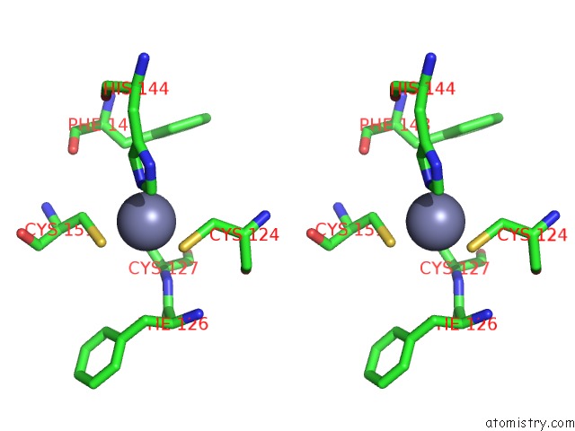
Stereo pair view
Mono view
Stereo pair view
A full contact list of Zinc with other atoms in the Zn binding
site number 1 of Structure of An Ml-Iap/Xiap Chimera Bound to A Peptidomimetic within 5.0Å range:
|
Zinc binding site 2 out of 2 in 2i3i
Go back to
Zinc binding site 2 out
of 2 in the Structure of An Ml-Iap/Xiap Chimera Bound to A Peptidomimetic
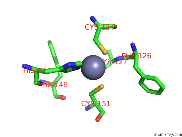
Mono view
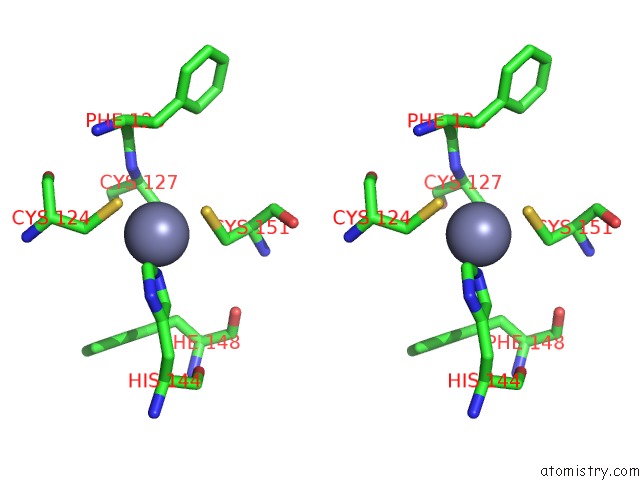
Stereo pair view
Mono view
Stereo pair view
A full contact list of Zinc with other atoms in the Zn binding
site number 2 of Structure of An Ml-Iap/Xiap Chimera Bound to A Peptidomimetic within 5.0Å range:
|
Reference:
K.Zobel,
L.Wang,
E.Varfolomeev,
M.C.Franklin,
L.O.Elliott,
H.J.Wallweber,
D.C.Okawa,
J.A.Flygare,
D.Vucic,
W.J.Fairbrother,
K.Deshayes.
Design, Synthesis, and Biological Activity of A Potent Smac Mimetic That Sensitizes Cancer Cells to Apoptosis By Antagonizing Iaps. Acs Chem.Biol. V. 1 525 2006.
ISSN: ISSN 1554-8929
PubMed: 17168540
DOI: 10.1021/CB600276Q
Page generated: Thu Oct 17 00:49:49 2024
ISSN: ISSN 1554-8929
PubMed: 17168540
DOI: 10.1021/CB600276Q
Last articles
Zn in 9MJ5Zn in 9HNW
Zn in 9G0L
Zn in 9FNE
Zn in 9DZN
Zn in 9E0I
Zn in 9D32
Zn in 9DAK
Zn in 8ZXC
Zn in 8ZUF