Zinc »
PDB 2hqk-2ibi »
2hue »
Zinc in PDB 2hue: Structure of the H3-H4 Chaperone ASF1 Bound to Histones H3 and H4
Protein crystallography data
The structure of Structure of the H3-H4 Chaperone ASF1 Bound to Histones H3 and H4, PDB code: 2hue
was solved by
C.M.English,
M.E.A.Churchill,
J.K.Tyler,
with X-Ray Crystallography technique. A brief refinement statistics is given in the table below:
| Resolution Low / High (Å) | 11.98 / 1.70 |
| Space group | P 31 2 1 |
| Cell size a, b, c (Å), α, β, γ (°) | 95.749, 95.749, 110.676, 90.00, 90.00, 120.00 |
| R / Rfree (%) | 20.9 / 23.9 |
Zinc Binding Sites:
The binding sites of Zinc atom in the Structure of the H3-H4 Chaperone ASF1 Bound to Histones H3 and H4
(pdb code 2hue). This binding sites where shown within
5.0 Angstroms radius around Zinc atom.
In total only one binding site of Zinc was determined in the Structure of the H3-H4 Chaperone ASF1 Bound to Histones H3 and H4, PDB code: 2hue:
In total only one binding site of Zinc was determined in the Structure of the H3-H4 Chaperone ASF1 Bound to Histones H3 and H4, PDB code: 2hue:
Zinc binding site 1 out of 1 in 2hue
Go back to
Zinc binding site 1 out
of 1 in the Structure of the H3-H4 Chaperone ASF1 Bound to Histones H3 and H4
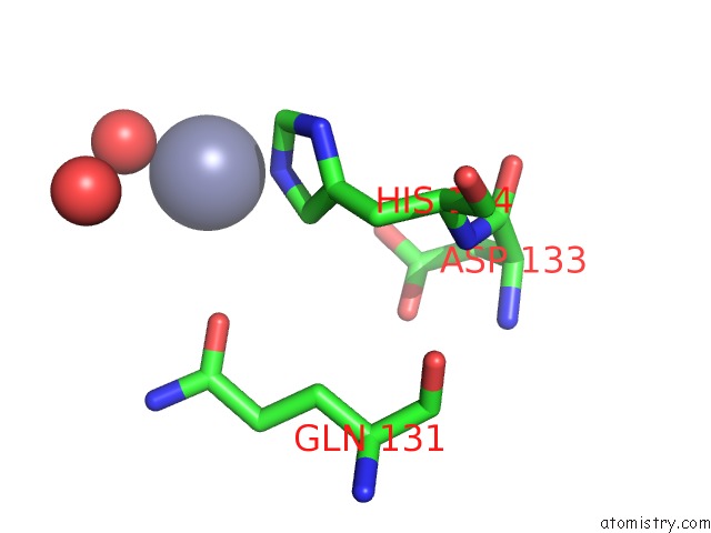
Mono view
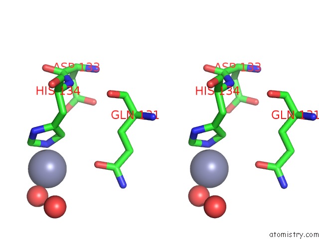
Stereo pair view
Mono view
Stereo pair view
A full contact list of Zinc with other atoms in the Zn binding
site number 1 of Structure of the H3-H4 Chaperone ASF1 Bound to Histones H3 and H4 within 5.0Å range:
|
Reference:
C.M.English,
M.W.Adkins,
J.J.Carson,
M.E.Churchill,
J.K.Tyler.
Structural Basis For the Histone Chaperone Activity of ASF1. Cell(Cambridge,Mass.) V. 127 495 2006.
ISSN: ISSN 0092-8674
PubMed: 17081973
DOI: 10.1016/J.CELL.2006.08.047
Page generated: Thu Oct 17 00:42:59 2024
ISSN: ISSN 0092-8674
PubMed: 17081973
DOI: 10.1016/J.CELL.2006.08.047
Last articles
Zn in 9MJ5Zn in 9HNW
Zn in 9G0L
Zn in 9FNE
Zn in 9DZN
Zn in 9E0I
Zn in 9D32
Zn in 9DAK
Zn in 8ZXC
Zn in 8ZUF