Zinc »
PDB 2hqk-2ibi »
2hqk »
Zinc in PDB 2hqk: Crystal Structure of A Monomeric Cyan Fluorescent Protein Derived From Clavularia
Protein crystallography data
The structure of Crystal Structure of A Monomeric Cyan Fluorescent Protein Derived From Clavularia, PDB code: 2hqk
was solved by
J.N.Henderson,
R.E.Campbell,
H.Ai,
S.J.Remington,
with X-Ray Crystallography technique. A brief refinement statistics is given in the table below:
| Resolution Low / High (Å) | 10.00 / 1.19 |
| Space group | C 1 2 1 |
| Cell size a, b, c (Å), α, β, γ (°) | 89.830, 38.020, 61.119, 90.00, 90.81, 90.00 |
| R / Rfree (%) | 14.9 / 20.6 |
Other elements in 2hqk:
The structure of Crystal Structure of A Monomeric Cyan Fluorescent Protein Derived From Clavularia also contains other interesting chemical elements:
| Chlorine | (Cl) | 1 atom |
Zinc Binding Sites:
The binding sites of Zinc atom in the Crystal Structure of A Monomeric Cyan Fluorescent Protein Derived From Clavularia
(pdb code 2hqk). This binding sites where shown within
5.0 Angstroms radius around Zinc atom.
In total 3 binding sites of Zinc where determined in the Crystal Structure of A Monomeric Cyan Fluorescent Protein Derived From Clavularia, PDB code: 2hqk:
Jump to Zinc binding site number: 1; 2; 3;
In total 3 binding sites of Zinc where determined in the Crystal Structure of A Monomeric Cyan Fluorescent Protein Derived From Clavularia, PDB code: 2hqk:
Jump to Zinc binding site number: 1; 2; 3;
Zinc binding site 1 out of 3 in 2hqk
Go back to
Zinc binding site 1 out
of 3 in the Crystal Structure of A Monomeric Cyan Fluorescent Protein Derived From Clavularia
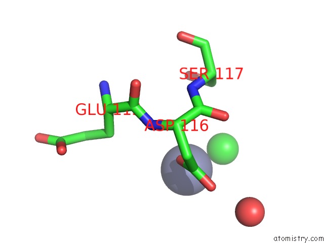
Mono view

Stereo pair view
Mono view
Stereo pair view
A full contact list of Zinc with other atoms in the Zn binding
site number 1 of Crystal Structure of A Monomeric Cyan Fluorescent Protein Derived From Clavularia within 5.0Å range:
|
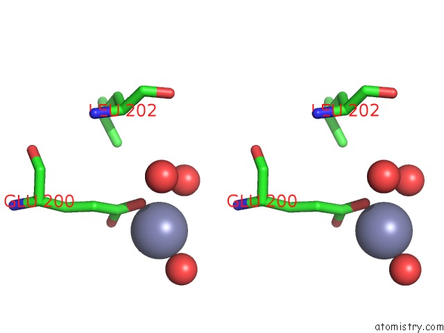
Zinc binding site 2 out of 3 in 2hqk
Go back to
Zinc binding site 2 out
of 3 in the Crystal Structure of A Monomeric Cyan Fluorescent Protein Derived From Clavularia

Mono view

Stereo pair view
Mono view
Stereo pair view
A full contact list of Zinc with other atoms in the Zn binding
site number 2 of Crystal Structure of A Monomeric Cyan Fluorescent Protein Derived From Clavularia within 5.0Å range:
|
Zinc binding site 3 out of 3 in 2hqk
Go back to
Zinc binding site 3 out
of 3 in the Crystal Structure of A Monomeric Cyan Fluorescent Protein Derived From Clavularia

Mono view
Stereo pair view
Mono view
Stereo pair view
A full contact list of Zinc with other atoms in the Zn binding
site number 3 of Crystal Structure of A Monomeric Cyan Fluorescent Protein Derived From Clavularia within 5.0Å range:
|
Reference:
H.W.Ai,
J.N.Henderson,
S.J.Remington,
R.E.Campbell.
Directed Evolution of A Monomeric, Bright and Photostable Version of Clavularia Cyan Fluorescent Protein: Structural Characterization and Applications in Fluorescence Imaging. Biochem.J. V. 400 531 2006.
ISSN: ISSN 0264-6021
PubMed: 16859491
DOI: 10.1042/BJ20060874
Page generated: Thu Oct 17 00:41:22 2024
ISSN: ISSN 0264-6021
PubMed: 16859491
DOI: 10.1042/BJ20060874
Last articles
Zn in 9MJ5Zn in 9HNW
Zn in 9G0L
Zn in 9FNE
Zn in 9DZN
Zn in 9E0I
Zn in 9D32
Zn in 9DAK
Zn in 8ZXC
Zn in 8ZUF