Zinc »
PDB 2hap-2hqh »
2hjh »
Zinc in PDB 2hjh: Crystal Structure of the SIR2 Deacetylase
Protein crystallography data
The structure of Crystal Structure of the SIR2 Deacetylase, PDB code: 2hjh
was solved by
B.E.Hall,
T.E.Ellenberger,
with X-Ray Crystallography technique. A brief refinement statistics is given in the table below:
| Resolution Low / High (Å) | 30.00 / 1.85 |
| Space group | P 1 21 1 |
| Cell size a, b, c (Å), α, β, γ (°) | 52.353, 89.566, 94.510, 90.00, 104.95, 90.00 |
| R / Rfree (%) | 17.1 / 21.5 |
Zinc Binding Sites:
The binding sites of Zinc atom in the Crystal Structure of the SIR2 Deacetylase
(pdb code 2hjh). This binding sites where shown within
5.0 Angstroms radius around Zinc atom.
In total 2 binding sites of Zinc where determined in the Crystal Structure of the SIR2 Deacetylase, PDB code: 2hjh:
Jump to Zinc binding site number: 1; 2;
In total 2 binding sites of Zinc where determined in the Crystal Structure of the SIR2 Deacetylase, PDB code: 2hjh:
Jump to Zinc binding site number: 1; 2;
Zinc binding site 1 out of 2 in 2hjh
Go back to
Zinc binding site 1 out
of 2 in the Crystal Structure of the SIR2 Deacetylase
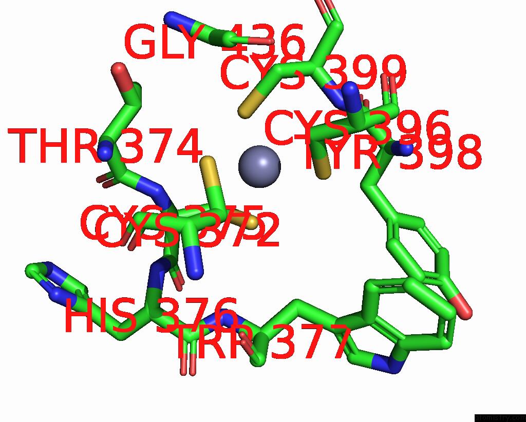
Mono view

Stereo pair view
Mono view
Stereo pair view
A full contact list of Zinc with other atoms in the Zn binding
site number 1 of Crystal Structure of the SIR2 Deacetylase within 5.0Å range:
|
Zinc binding site 2 out of 2 in 2hjh
Go back to
Zinc binding site 2 out
of 2 in the Crystal Structure of the SIR2 Deacetylase

Mono view

Stereo pair view
Mono view
Stereo pair view
A full contact list of Zinc with other atoms in the Zn binding
site number 2 of Crystal Structure of the SIR2 Deacetylase within 5.0Å range:
|
Reference:
B.E.Hall,
J.R.Buchberger,
S.A.Gerber,
A.L.B.Ambrosio,
S.P.Gygi,
D.Filman,
D.Moazed,
T.Ellenberger.
Autoregulation of the Yeast SIR2 Deacetylase By Reaction and Trapping of A Pseudosubstrate Motif in the Active Site To Be Published.
Page generated: Thu Oct 17 00:39:41 2024
Last articles
Al in 8UQNAl in 8TSI
Al in 8T0L
Al in 8SHO
Al in 8SHP
Al in 8SHN
Al in 8SHL
Al in 8SHE
Al in 8SHG
Al in 8SHF