Zinc »
PDB 2hap-2hqh »
2hf8 »
Zinc in PDB 2hf8: Crystal Structure of Hypb From Methanocaldococcus Jannaschii in the Triphosphate Form, in Complex with Zinc
Protein crystallography data
The structure of Crystal Structure of Hypb From Methanocaldococcus Jannaschii in the Triphosphate Form, in Complex with Zinc, PDB code: 2hf8
was solved by
R.Gasper,
A.Scrima,
A.Wittinghofer,
with X-Ray Crystallography technique. A brief refinement statistics is given in the table below:
| Resolution Low / High (Å) | 19.92 / 2.10 |
| Space group | P 21 21 21 |
| Cell size a, b, c (Å), α, β, γ (°) | 43.120, 68.130, 155.990, 90.00, 90.00, 90.00 |
| R / Rfree (%) | 20.1 / 24.2 |
Other elements in 2hf8:
The structure of Crystal Structure of Hypb From Methanocaldococcus Jannaschii in the Triphosphate Form, in Complex with Zinc also contains other interesting chemical elements:
| Magnesium | (Mg) | 2 atoms |
Zinc Binding Sites:
The binding sites of Zinc atom in the Crystal Structure of Hypb From Methanocaldococcus Jannaschii in the Triphosphate Form, in Complex with Zinc
(pdb code 2hf8). This binding sites where shown within
5.0 Angstroms radius around Zinc atom.
In total 4 binding sites of Zinc where determined in the Crystal Structure of Hypb From Methanocaldococcus Jannaschii in the Triphosphate Form, in Complex with Zinc, PDB code: 2hf8:
Jump to Zinc binding site number: 1; 2; 3; 4;
In total 4 binding sites of Zinc where determined in the Crystal Structure of Hypb From Methanocaldococcus Jannaschii in the Triphosphate Form, in Complex with Zinc, PDB code: 2hf8:
Jump to Zinc binding site number: 1; 2; 3; 4;
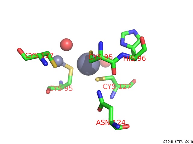
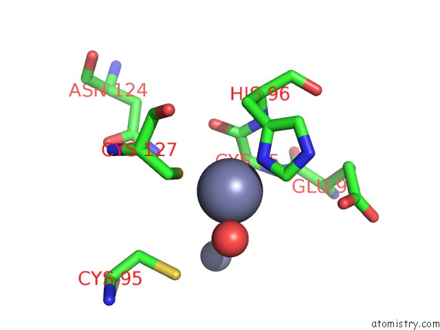
Zinc binding site 1 out of 4 in 2hf8
Go back to
Zinc binding site 1 out
of 4 in the Crystal Structure of Hypb From Methanocaldococcus Jannaschii in the Triphosphate Form, in Complex with Zinc
Mono view
Stereo pair view
Mono view
Stereo pair view
A full contact list of Zinc with other atoms in the Zn binding
site number 1 of Crystal Structure of Hypb From Methanocaldococcus Jannaschii in the Triphosphate Form, in Complex with Zinc within 5.0Å range:
|
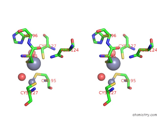
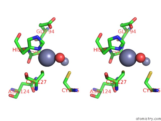
Zinc binding site 2 out of 4 in 2hf8
Go back to
Zinc binding site 2 out
of 4 in the Crystal Structure of Hypb From Methanocaldococcus Jannaschii in the Triphosphate Form, in Complex with Zinc
Mono view
Stereo pair view
Mono view
Stereo pair view
A full contact list of Zinc with other atoms in the Zn binding
site number 2 of Crystal Structure of Hypb From Methanocaldococcus Jannaschii in the Triphosphate Form, in Complex with Zinc within 5.0Å range:
|
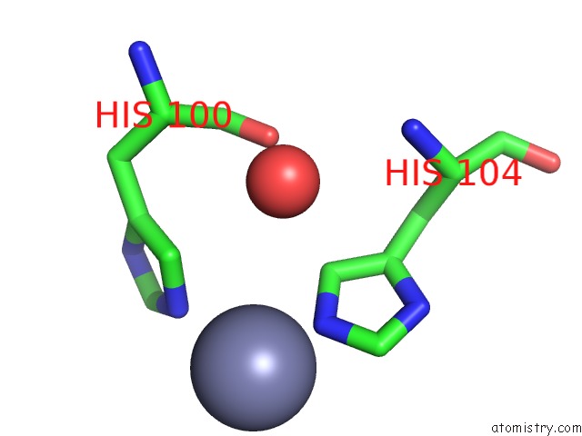
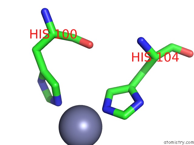
Zinc binding site 3 out of 4 in 2hf8
Go back to
Zinc binding site 3 out
of 4 in the Crystal Structure of Hypb From Methanocaldococcus Jannaschii in the Triphosphate Form, in Complex with Zinc
Mono view
Stereo pair view
Mono view
Stereo pair view
A full contact list of Zinc with other atoms in the Zn binding
site number 3 of Crystal Structure of Hypb From Methanocaldococcus Jannaschii in the Triphosphate Form, in Complex with Zinc within 5.0Å range:
|
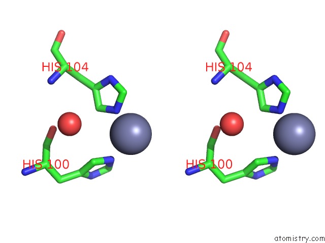
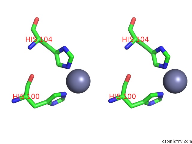
Zinc binding site 4 out of 4 in 2hf8
Go back to
Zinc binding site 4 out
of 4 in the Crystal Structure of Hypb From Methanocaldococcus Jannaschii in the Triphosphate Form, in Complex with Zinc
Mono view
Stereo pair view
Mono view
Stereo pair view
A full contact list of Zinc with other atoms in the Zn binding
site number 4 of Crystal Structure of Hypb From Methanocaldococcus Jannaschii in the Triphosphate Form, in Complex with Zinc within 5.0Å range:
|
Reference:
R.Gasper,
A.Scrima,
A.Wittinghofer.
Structural Insights Into Hypb, A Gtp-Binding Protein That Regulates Metal Binding. J.Biol.Chem. V. 281 27492 2006.
ISSN: ISSN 0021-9258
PubMed: 16807243
DOI: 10.1074/JBC.M600809200
Page generated: Thu Oct 17 00:38:05 2024
ISSN: ISSN 0021-9258
PubMed: 16807243
DOI: 10.1074/JBC.M600809200
Last articles
Zn in 9MJ5Zn in 9HNW
Zn in 9G0L
Zn in 9FNE
Zn in 9DZN
Zn in 9E0I
Zn in 9D32
Zn in 9DAK
Zn in 8ZXC
Zn in 8ZUF