Zinc »
PDB 2geh-2gzg »
2gu2 »
Zinc in PDB 2gu2: Crystal Structure of An Aspartoacylase From Rattus Norvegicus
Enzymatic activity of Crystal Structure of An Aspartoacylase From Rattus Norvegicus
All present enzymatic activity of Crystal Structure of An Aspartoacylase From Rattus Norvegicus:
3.5.1.15;
3.5.1.15;
Protein crystallography data
The structure of Crystal Structure of An Aspartoacylase From Rattus Norvegicus, PDB code: 2gu2
was solved by
E.Bitto,
G.E.Wesenberg,
G.N.Phillips Jr.,
C.A.Bingman,
Center Foreukaryotic Structural Genomics (Cesg),
with X-Ray Crystallography technique. A brief refinement statistics is given in the table below:
| Resolution Low / High (Å) | 41.75 / 1.81 |
| Space group | C 1 2 1 |
| Cell size a, b, c (Å), α, β, γ (°) | 92.581, 135.778, 54.033, 90.00, 101.49, 90.00 |
| R / Rfree (%) | 14.9 / 19.4 |
Zinc Binding Sites:
The binding sites of Zinc atom in the Crystal Structure of An Aspartoacylase From Rattus Norvegicus
(pdb code 2gu2). This binding sites where shown within
5.0 Angstroms radius around Zinc atom.
In total 2 binding sites of Zinc where determined in the Crystal Structure of An Aspartoacylase From Rattus Norvegicus, PDB code: 2gu2:
Jump to Zinc binding site number: 1; 2;
In total 2 binding sites of Zinc where determined in the Crystal Structure of An Aspartoacylase From Rattus Norvegicus, PDB code: 2gu2:
Jump to Zinc binding site number: 1; 2;
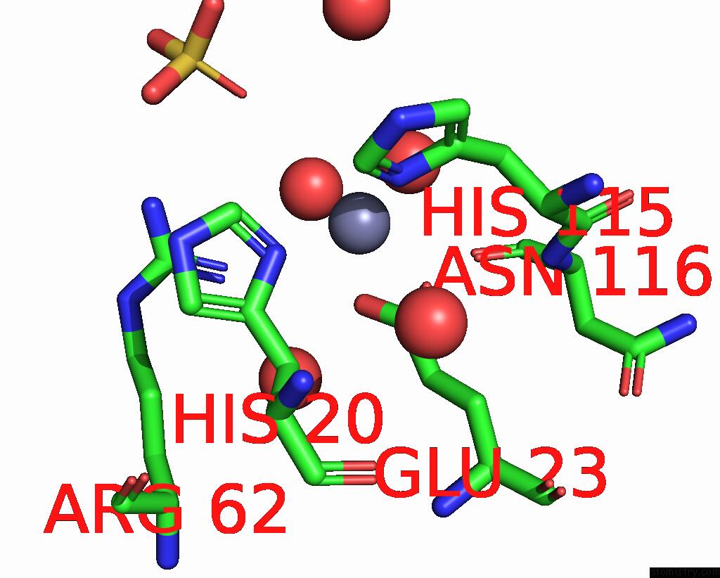
Zinc binding site 1 out of 2 in 2gu2
Go back to
Zinc binding site 1 out
of 2 in the Crystal Structure of An Aspartoacylase From Rattus Norvegicus

Mono view
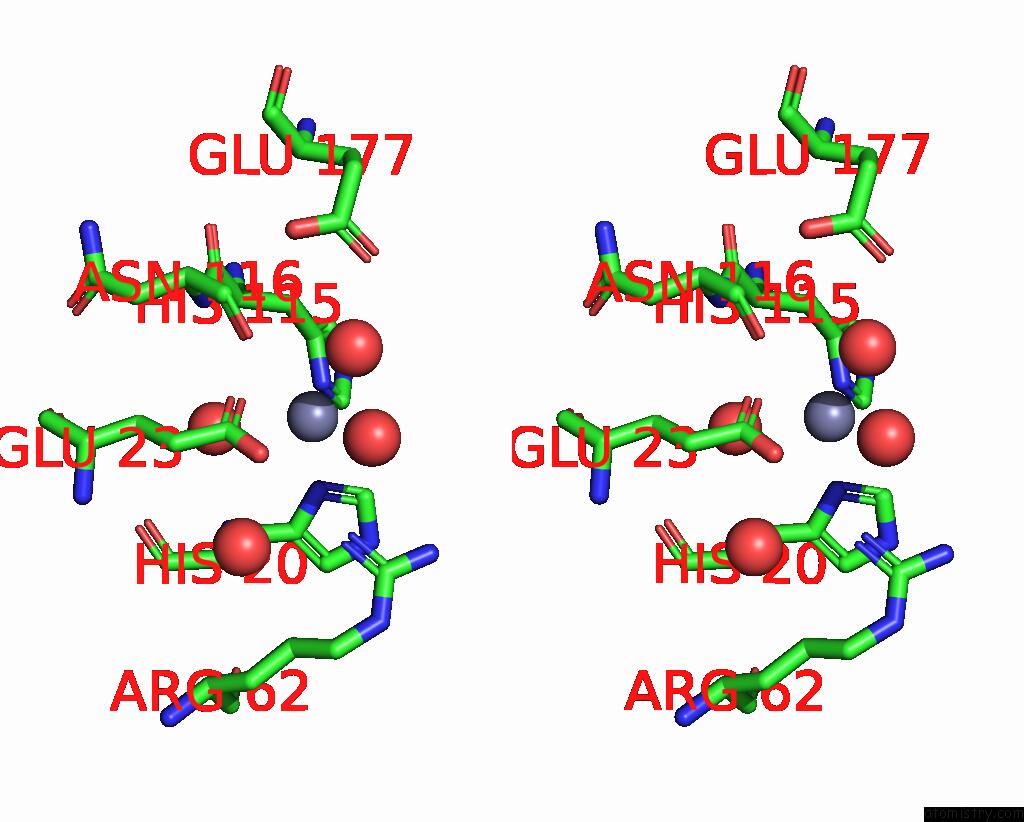
Stereo pair view
Mono view
Stereo pair view
A full contact list of Zinc with other atoms in the Zn binding
site number 1 of Crystal Structure of An Aspartoacylase From Rattus Norvegicus within 5.0Å range:
|
Zinc binding site 2 out of 2 in 2gu2
Go back to
Zinc binding site 2 out
of 2 in the Crystal Structure of An Aspartoacylase From Rattus Norvegicus
Mono view
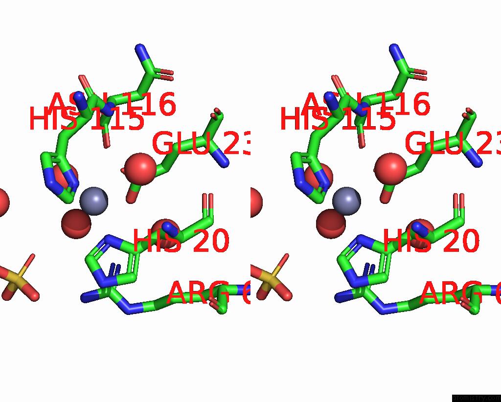
Stereo pair view
Mono view
Stereo pair view
A full contact list of Zinc with other atoms in the Zn binding
site number 2 of Crystal Structure of An Aspartoacylase From Rattus Norvegicus within 5.0Å range:
|
Reference:
E.Bitto,
C.A.Bingman,
G.E.Wesenberg,
J.G.Mccoy,
G.N.Phillips.
Structure of Aspartoacylase, the Brain Enzyme Impaired in Canavan Disease. Proc.Natl.Acad.Sci.Usa V. 104 456 2007.
ISSN: ISSN 0027-8424
PubMed: 17194761
DOI: 10.1073/PNAS.0607817104
Page generated: Thu Oct 17 00:24:34 2024
ISSN: ISSN 0027-8424
PubMed: 17194761
DOI: 10.1073/PNAS.0607817104
Last articles
Zn in 9MJ5Zn in 9HNW
Zn in 9G0L
Zn in 9FNE
Zn in 9DZN
Zn in 9E0I
Zn in 9D32
Zn in 9DAK
Zn in 8ZXC
Zn in 8ZUF