Zinc »
PDB 2geh-2gzg »
2gmw »
Zinc in PDB 2gmw: Crystal Structure of D,D-Heptose 1.7-Bisphosphate Phosphatase From E. Coli.
Protein crystallography data
The structure of Crystal Structure of D,D-Heptose 1.7-Bisphosphate Phosphatase From E. Coli., PDB code: 2gmw
was solved by
K.Zhang,
G.Deleon,
G.D.Wright,
M.S.Junop,
with X-Ray Crystallography technique. A brief refinement statistics is given in the table below:
| Resolution Low / High (Å) | 25.00 / 1.50 |
| Space group | P 21 21 21 |
| Cell size a, b, c (Å), α, β, γ (°) | 51.904, 63.977, 103.324, 90.00, 90.00, 90.00 |
| R / Rfree (%) | 16.3 / 21.2 |
Zinc Binding Sites:
The binding sites of Zinc atom in the Crystal Structure of D,D-Heptose 1.7-Bisphosphate Phosphatase From E. Coli.
(pdb code 2gmw). This binding sites where shown within
5.0 Angstroms radius around Zinc atom.
In total 2 binding sites of Zinc where determined in the Crystal Structure of D,D-Heptose 1.7-Bisphosphate Phosphatase From E. Coli., PDB code: 2gmw:
Jump to Zinc binding site number: 1; 2;
In total 2 binding sites of Zinc where determined in the Crystal Structure of D,D-Heptose 1.7-Bisphosphate Phosphatase From E. Coli., PDB code: 2gmw:
Jump to Zinc binding site number: 1; 2;
Zinc binding site 1 out of 2 in 2gmw
Go back to
Zinc binding site 1 out
of 2 in the Crystal Structure of D,D-Heptose 1.7-Bisphosphate Phosphatase From E. Coli.
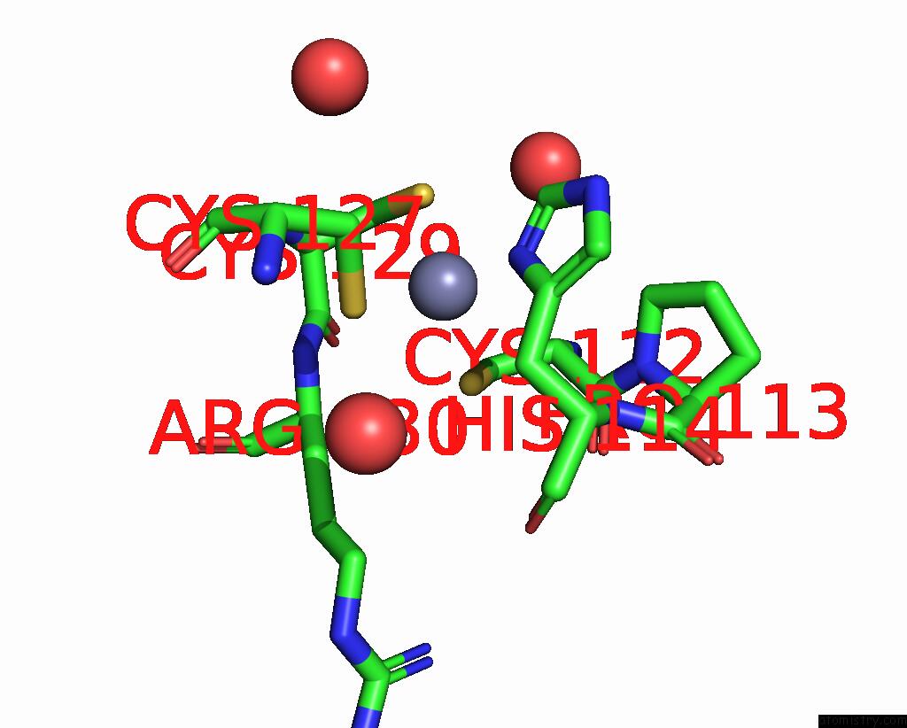
Mono view
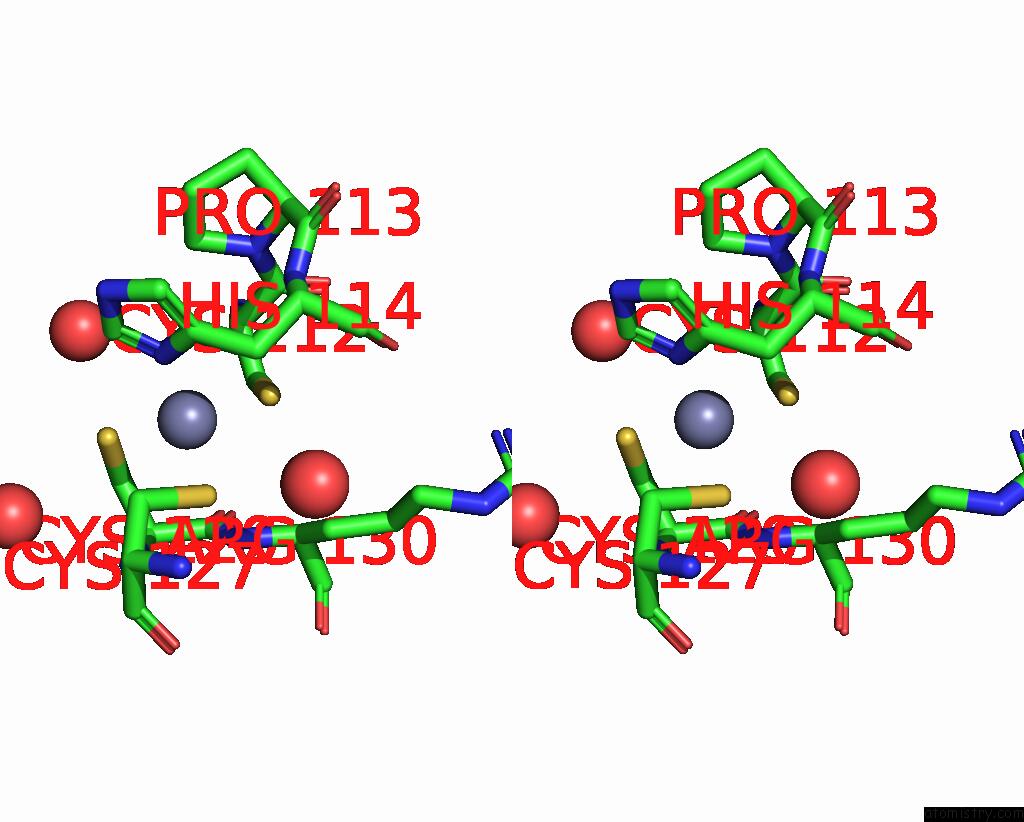
Stereo pair view
Mono view
Stereo pair view
A full contact list of Zinc with other atoms in the Zn binding
site number 1 of Crystal Structure of D,D-Heptose 1.7-Bisphosphate Phosphatase From E. Coli. within 5.0Å range:
|
Zinc binding site 2 out of 2 in 2gmw
Go back to
Zinc binding site 2 out
of 2 in the Crystal Structure of D,D-Heptose 1.7-Bisphosphate Phosphatase From E. Coli.
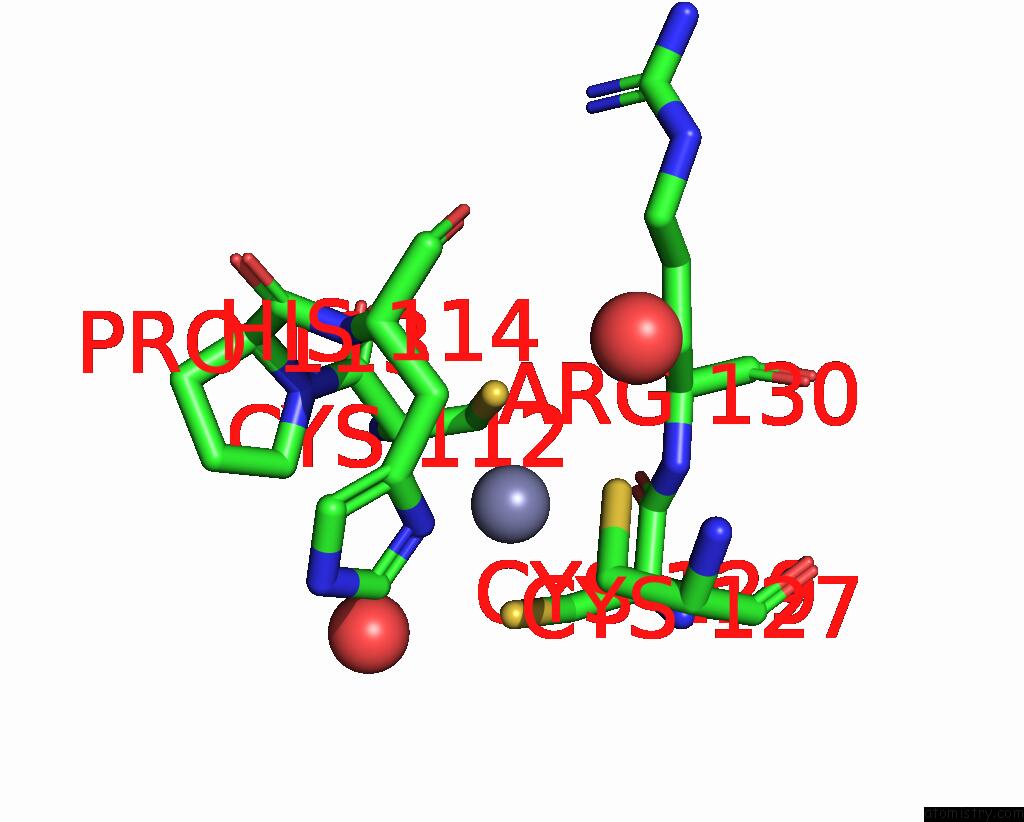
Mono view
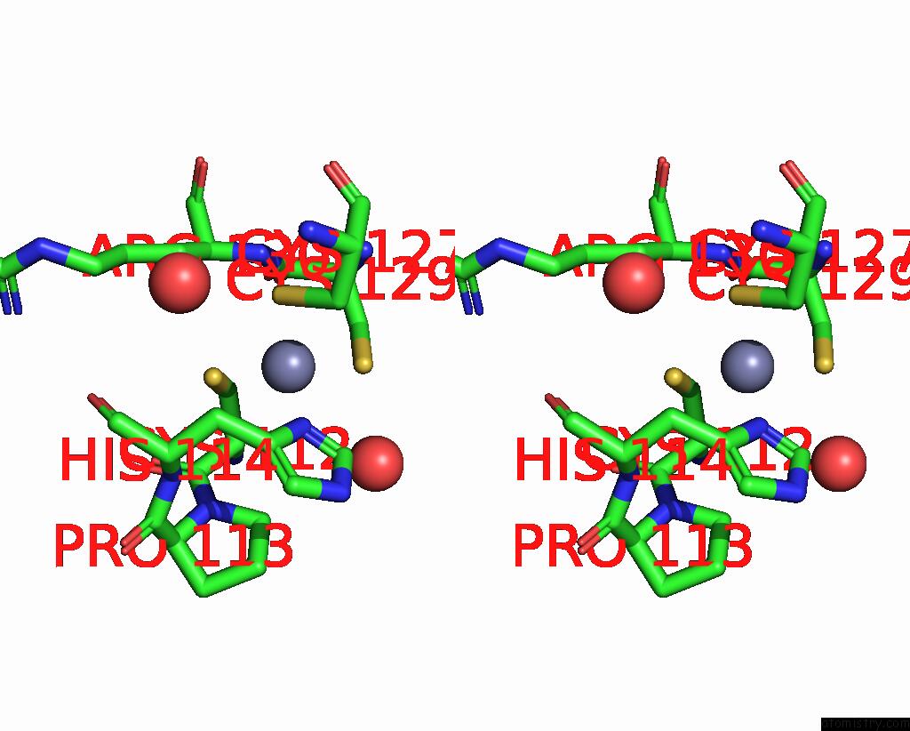
Stereo pair view
Mono view
Stereo pair view
A full contact list of Zinc with other atoms in the Zn binding
site number 2 of Crystal Structure of D,D-Heptose 1.7-Bisphosphate Phosphatase From E. Coli. within 5.0Å range:
|
Reference:
P.L.Taylor,
S.Sugiman-Marangos,
K.Zhang,
M.A.Valvano,
G.D.Wright,
M.S.Junop.
Structural and Kinetic Characterization of the Lps Biosynthetic Enzyme D-Alpha,Beta-D-Heptose-1,7-Bisphosphate Phosphatase (Gmhb) From Escherichia Coli. Biochemistry V. 49 1033 2010.
ISSN: ISSN 0006-2960
PubMed: 20050699
DOI: 10.1021/BI901780J
Page generated: Thu Oct 17 00:20:43 2024
ISSN: ISSN 0006-2960
PubMed: 20050699
DOI: 10.1021/BI901780J
Last articles
Zn in 9MJ5Zn in 9HNW
Zn in 9G0L
Zn in 9FNE
Zn in 9DZN
Zn in 9E0I
Zn in 9D32
Zn in 9DAK
Zn in 8ZXC
Zn in 8ZUF