Zinc »
PDB 2geh-2gzg »
2geq »
Zinc in PDB 2geq: Crystal Structure of A P53 Core Dimer Bound to Dna
Protein crystallography data
The structure of Crystal Structure of A P53 Core Dimer Bound to Dna, PDB code: 2geq
was solved by
W.C.Ho,
M.X.Fitzgerald,
R.Marmorstein,
with X-Ray Crystallography technique. A brief refinement statistics is given in the table below:
| Resolution Low / High (Å) | 47.15 / 2.30 |
| Space group | P 21 21 21 |
| Cell size a, b, c (Å), α, β, γ (°) | 44.755, 94.290, 119.822, 90.00, 90.00, 90.00 |
| R / Rfree (%) | 20.2 / 23.8 |
Zinc Binding Sites:
The binding sites of Zinc atom in the Crystal Structure of A P53 Core Dimer Bound to Dna
(pdb code 2geq). This binding sites where shown within
5.0 Angstroms radius around Zinc atom.
In total 2 binding sites of Zinc where determined in the Crystal Structure of A P53 Core Dimer Bound to Dna, PDB code: 2geq:
Jump to Zinc binding site number: 1; 2;
In total 2 binding sites of Zinc where determined in the Crystal Structure of A P53 Core Dimer Bound to Dna, PDB code: 2geq:
Jump to Zinc binding site number: 1; 2;
Zinc binding site 1 out of 2 in 2geq
Go back to
Zinc binding site 1 out
of 2 in the Crystal Structure of A P53 Core Dimer Bound to Dna
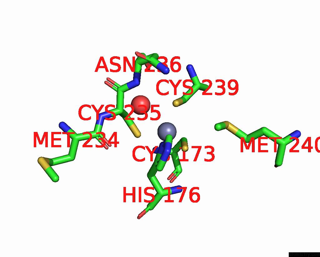
Mono view
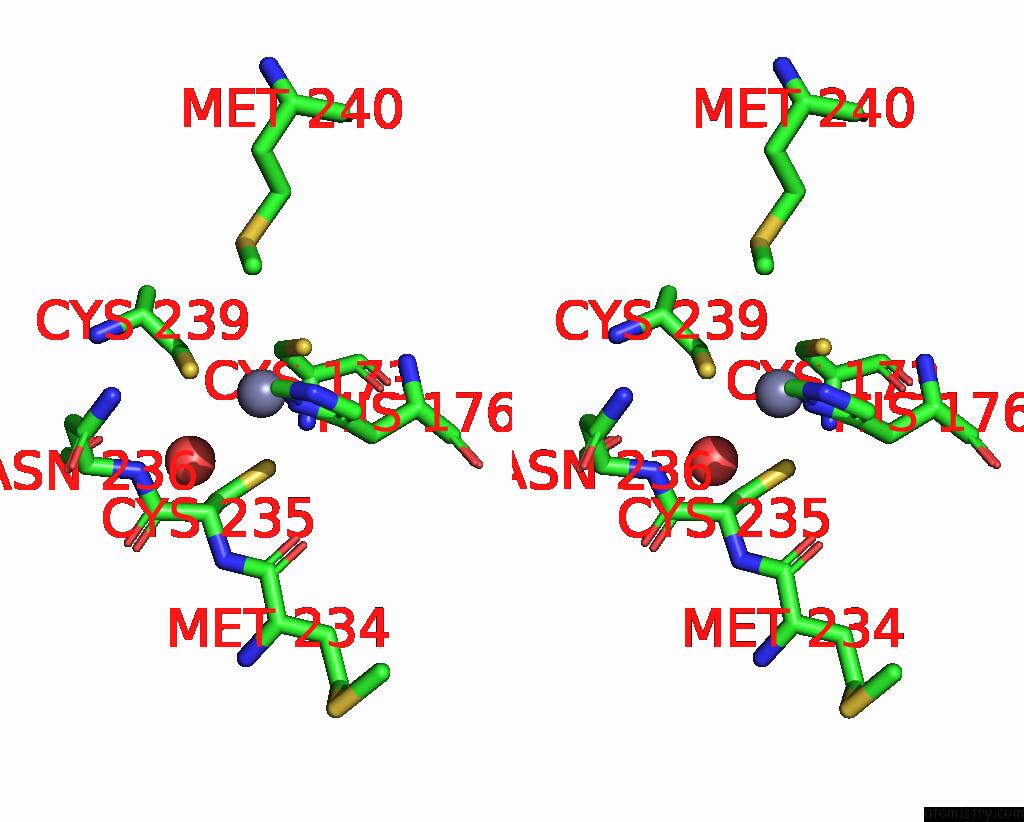
Stereo pair view
Mono view
Stereo pair view
A full contact list of Zinc with other atoms in the Zn binding
site number 1 of Crystal Structure of A P53 Core Dimer Bound to Dna within 5.0Å range:
|
Zinc binding site 2 out of 2 in 2geq
Go back to
Zinc binding site 2 out
of 2 in the Crystal Structure of A P53 Core Dimer Bound to Dna

Mono view
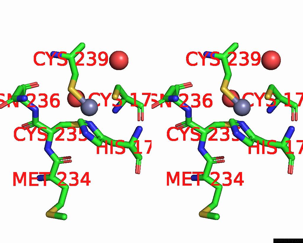
Stereo pair view
Mono view
Stereo pair view
A full contact list of Zinc with other atoms in the Zn binding
site number 2 of Crystal Structure of A P53 Core Dimer Bound to Dna within 5.0Å range:
|
Reference:
W.C.Ho,
M.X.Fitzgerald,
R.Marmorstein.
Structure of the P53 Core Domain Dimer Bound to Dna. J.Biol.Chem. V. 281 20494 2006.
ISSN: ISSN 0021-9258
PubMed: 16717092
DOI: 10.1074/JBC.M603634200
Page generated: Thu Oct 17 00:17:42 2024
ISSN: ISSN 0021-9258
PubMed: 16717092
DOI: 10.1074/JBC.M603634200
Last articles
Zn in 9MJ5Zn in 9HNW
Zn in 9G0L
Zn in 9FNE
Zn in 9DZN
Zn in 9E0I
Zn in 9D32
Zn in 9DAK
Zn in 8ZXC
Zn in 8ZUF