Zinc »
PDB 2g3f-2gda »
2g54 »
Zinc in PDB 2g54: Crystal Structure of Zn-Bound Human Insulin-Degrading Enzyme in Complex with Insulin B Chain
Enzymatic activity of Crystal Structure of Zn-Bound Human Insulin-Degrading Enzyme in Complex with Insulin B Chain
All present enzymatic activity of Crystal Structure of Zn-Bound Human Insulin-Degrading Enzyme in Complex with Insulin B Chain:
3.4.24.56;
3.4.24.56;
Protein crystallography data
The structure of Crystal Structure of Zn-Bound Human Insulin-Degrading Enzyme in Complex with Insulin B Chain, PDB code: 2g54
was solved by
Y.Shen,
W.-J.Tang,
with X-Ray Crystallography technique. A brief refinement statistics is given in the table below:
| Resolution Low / High (Å) | 28.03 / 2.25 |
| Space group | P 65 |
| Cell size a, b, c (Å), α, β, γ (°) | 262.528, 262.528, 90.503, 90.00, 90.00, 120.00 |
| R / Rfree (%) | 20.6 / 23.3 |
Zinc Binding Sites:
The binding sites of Zinc atom in the Crystal Structure of Zn-Bound Human Insulin-Degrading Enzyme in Complex with Insulin B Chain
(pdb code 2g54). This binding sites where shown within
5.0 Angstroms radius around Zinc atom.
In total 2 binding sites of Zinc where determined in the Crystal Structure of Zn-Bound Human Insulin-Degrading Enzyme in Complex with Insulin B Chain, PDB code: 2g54:
Jump to Zinc binding site number: 1; 2;
In total 2 binding sites of Zinc where determined in the Crystal Structure of Zn-Bound Human Insulin-Degrading Enzyme in Complex with Insulin B Chain, PDB code: 2g54:
Jump to Zinc binding site number: 1; 2;
Zinc binding site 1 out of 2 in 2g54
Go back to
Zinc binding site 1 out
of 2 in the Crystal Structure of Zn-Bound Human Insulin-Degrading Enzyme in Complex with Insulin B Chain
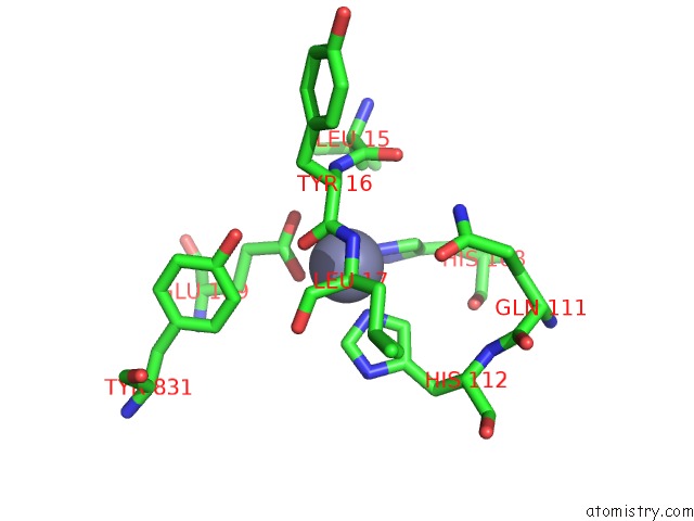
Mono view
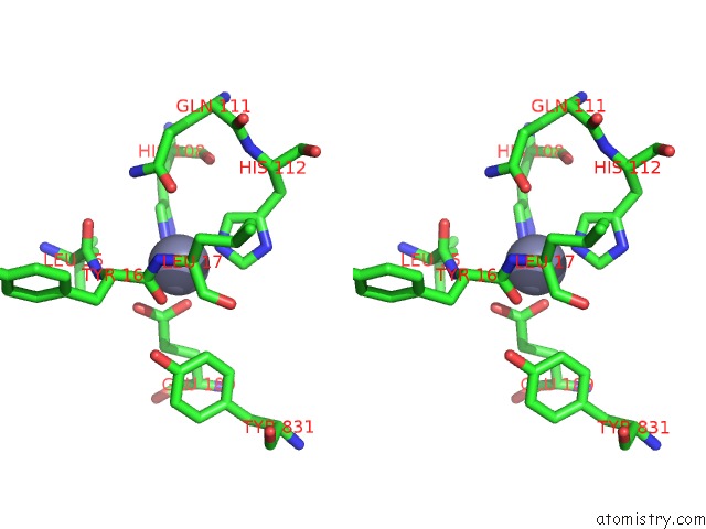
Stereo pair view
Mono view
Stereo pair view
A full contact list of Zinc with other atoms in the Zn binding
site number 1 of Crystal Structure of Zn-Bound Human Insulin-Degrading Enzyme in Complex with Insulin B Chain within 5.0Å range:
|
Zinc binding site 2 out of 2 in 2g54
Go back to
Zinc binding site 2 out
of 2 in the Crystal Structure of Zn-Bound Human Insulin-Degrading Enzyme in Complex with Insulin B Chain
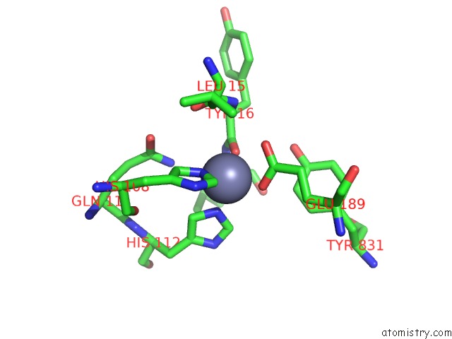
Mono view
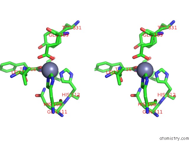
Stereo pair view
Mono view
Stereo pair view
A full contact list of Zinc with other atoms in the Zn binding
site number 2 of Crystal Structure of Zn-Bound Human Insulin-Degrading Enzyme in Complex with Insulin B Chain within 5.0Å range:
|
Reference:
Y.Shen,
A.Joachimiak,
M.R.Rosner,
W.J.Tang.
Structures of Human Insulin-Degrading Enzyme Reveal A New Substrate Recognition Mechanism. Nature V. 443 870 2006.
ISSN: ISSN 0028-0836
PubMed: 17051221
DOI: 10.1038/NATURE05143
Page generated: Thu Oct 17 00:01:28 2024
ISSN: ISSN 0028-0836
PubMed: 17051221
DOI: 10.1038/NATURE05143
Last articles
Zn in 9MJ5Zn in 9HNW
Zn in 9G0L
Zn in 9FNE
Zn in 9DZN
Zn in 9E0I
Zn in 9D32
Zn in 9DAK
Zn in 8ZXC
Zn in 8ZUF