Zinc »
PDB 2fos-2g2p »
2fvk »
Zinc in PDB 2fvk: Crystal Structure of Dihydropyrimidinase From Saccharomyces Kluyveri in Complex with the Substrate Dihydrouracil
Enzymatic activity of Crystal Structure of Dihydropyrimidinase From Saccharomyces Kluyveri in Complex with the Substrate Dihydrouracil
All present enzymatic activity of Crystal Structure of Dihydropyrimidinase From Saccharomyces Kluyveri in Complex with the Substrate Dihydrouracil:
3.5.2.2;
3.5.2.2;
Protein crystallography data
The structure of Crystal Structure of Dihydropyrimidinase From Saccharomyces Kluyveri in Complex with the Substrate Dihydrouracil, PDB code: 2fvk
was solved by
D.Dobritzsch,
B.Lohkamp,
with X-Ray Crystallography technique. A brief refinement statistics is given in the table below:
| Resolution Low / High (Å) | 54.23 / 2.40 |
| Space group | P 1 21 1 |
| Cell size a, b, c (Å), α, β, γ (°) | 90.130, 71.600, 161.890, 90.00, 91.40, 90.00 |
| R / Rfree (%) | 17.8 / 23.4 |
Zinc Binding Sites:
The binding sites of Zinc atom in the Crystal Structure of Dihydropyrimidinase From Saccharomyces Kluyveri in Complex with the Substrate Dihydrouracil
(pdb code 2fvk). This binding sites where shown within
5.0 Angstroms radius around Zinc atom.
In total 8 binding sites of Zinc where determined in the Crystal Structure of Dihydropyrimidinase From Saccharomyces Kluyveri in Complex with the Substrate Dihydrouracil, PDB code: 2fvk:
Jump to Zinc binding site number: 1; 2; 3; 4; 5; 6; 7; 8;
In total 8 binding sites of Zinc where determined in the Crystal Structure of Dihydropyrimidinase From Saccharomyces Kluyveri in Complex with the Substrate Dihydrouracil, PDB code: 2fvk:
Jump to Zinc binding site number: 1; 2; 3; 4; 5; 6; 7; 8;
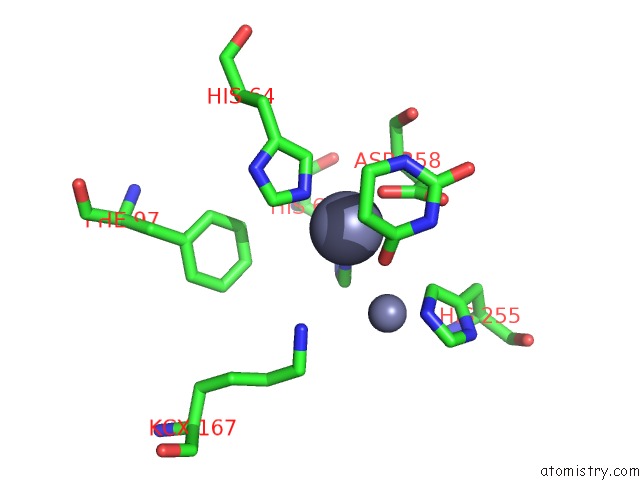
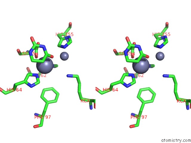
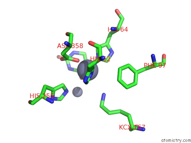
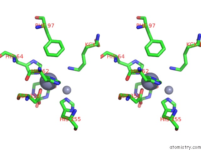
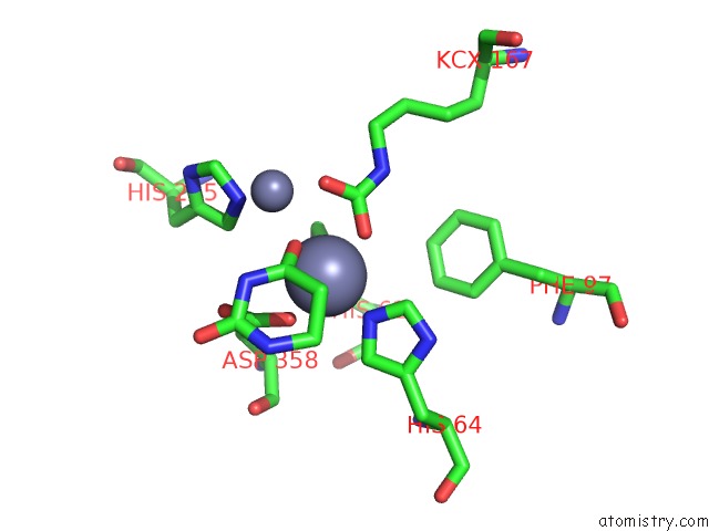
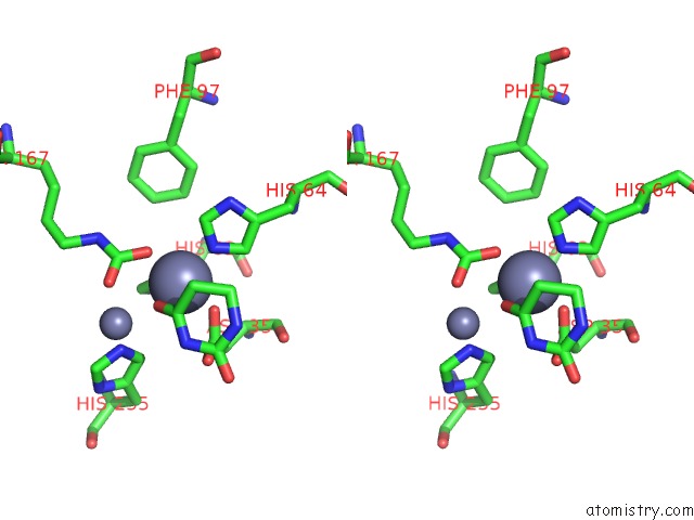
Zinc binding site 1 out of 8 in 2fvk
Go back to
Zinc binding site 1 out
of 8 in the Crystal Structure of Dihydropyrimidinase From Saccharomyces Kluyveri in Complex with the Substrate Dihydrouracil
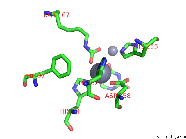
Mono view
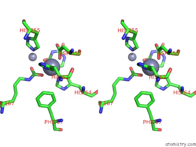
Stereo pair view
Mono view
Stereo pair view
A full contact list of Zinc with other atoms in the Zn binding
site number 1 of Crystal Structure of Dihydropyrimidinase From Saccharomyces Kluyveri in Complex with the Substrate Dihydrouracil within 5.0Å range:
|
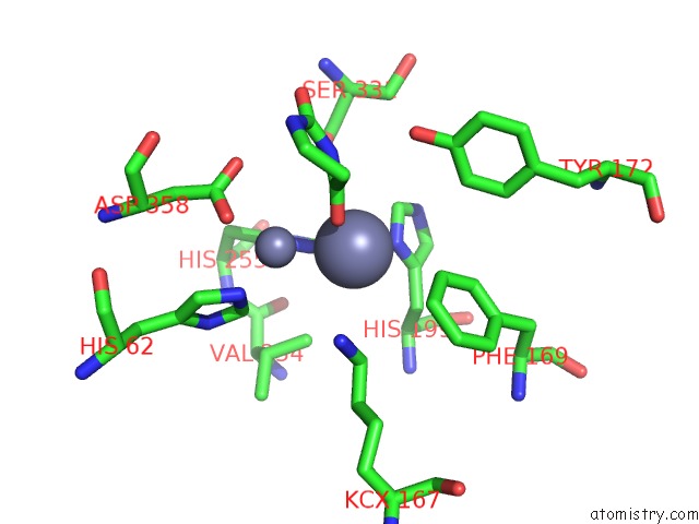
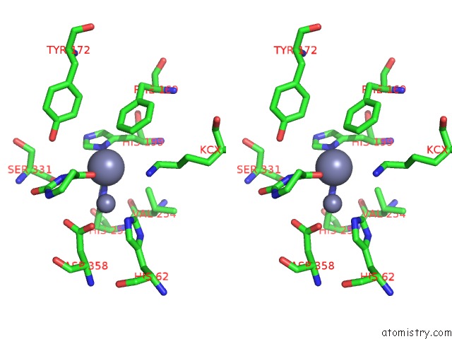
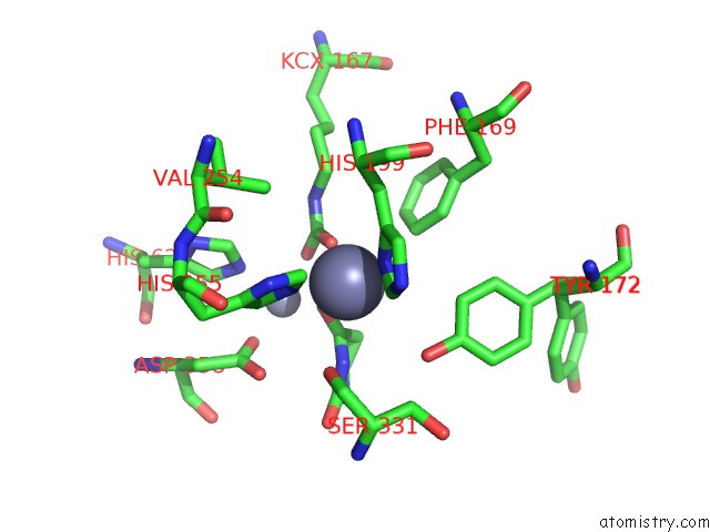
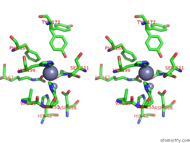
Zinc binding site 2 out of 8 in 2fvk
Go back to
Zinc binding site 2 out
of 8 in the Crystal Structure of Dihydropyrimidinase From Saccharomyces Kluyveri in Complex with the Substrate Dihydrouracil
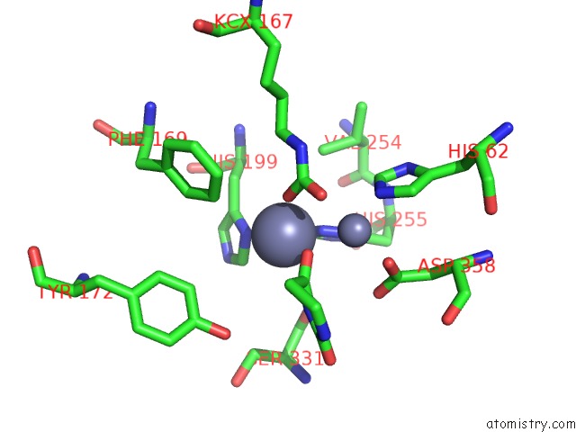
Mono view
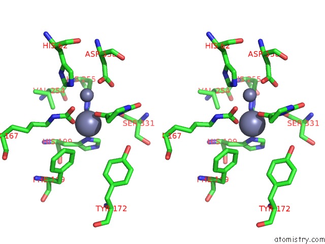
Stereo pair view
Mono view
Stereo pair view
A full contact list of Zinc with other atoms in the Zn binding
site number 2 of Crystal Structure of Dihydropyrimidinase From Saccharomyces Kluyveri in Complex with the Substrate Dihydrouracil within 5.0Å range:
|
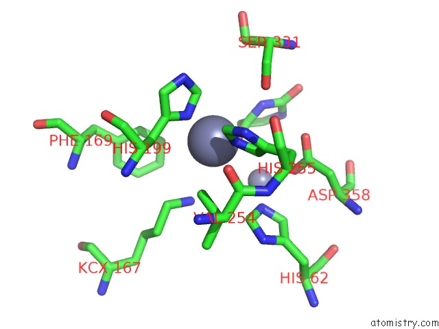
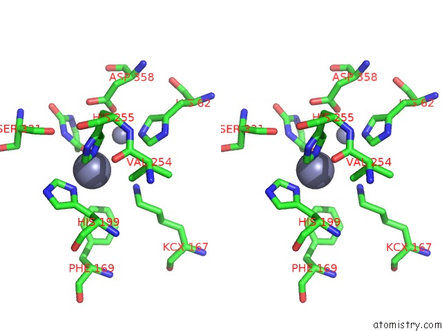
Zinc binding site 3 out of 8 in 2fvk
Go back to
Zinc binding site 3 out
of 8 in the Crystal Structure of Dihydropyrimidinase From Saccharomyces Kluyveri in Complex with the Substrate Dihydrouracil
Mono view
Stereo pair view
Mono view
Stereo pair view
A full contact list of Zinc with other atoms in the Zn binding
site number 3 of Crystal Structure of Dihydropyrimidinase From Saccharomyces Kluyveri in Complex with the Substrate Dihydrouracil within 5.0Å range:
|
Zinc binding site 4 out of 8 in 2fvk
Go back to
Zinc binding site 4 out
of 8 in the Crystal Structure of Dihydropyrimidinase From Saccharomyces Kluyveri in Complex with the Substrate Dihydrouracil
Mono view
Stereo pair view
Mono view
Stereo pair view
A full contact list of Zinc with other atoms in the Zn binding
site number 4 of Crystal Structure of Dihydropyrimidinase From Saccharomyces Kluyveri in Complex with the Substrate Dihydrouracil within 5.0Å range:
|
Zinc binding site 5 out of 8 in 2fvk
Go back to
Zinc binding site 5 out
of 8 in the Crystal Structure of Dihydropyrimidinase From Saccharomyces Kluyveri in Complex with the Substrate Dihydrouracil
Mono view
Stereo pair view
Mono view
Stereo pair view
A full contact list of Zinc with other atoms in the Zn binding
site number 5 of Crystal Structure of Dihydropyrimidinase From Saccharomyces Kluyveri in Complex with the Substrate Dihydrouracil within 5.0Å range:
|
Zinc binding site 6 out of 8 in 2fvk
Go back to
Zinc binding site 6 out
of 8 in the Crystal Structure of Dihydropyrimidinase From Saccharomyces Kluyveri in Complex with the Substrate Dihydrouracil
Mono view
Stereo pair view
Mono view
Stereo pair view
A full contact list of Zinc with other atoms in the Zn binding
site number 6 of Crystal Structure of Dihydropyrimidinase From Saccharomyces Kluyveri in Complex with the Substrate Dihydrouracil within 5.0Å range:
|
Zinc binding site 7 out of 8 in 2fvk
Go back to
Zinc binding site 7 out
of 8 in the Crystal Structure of Dihydropyrimidinase From Saccharomyces Kluyveri in Complex with the Substrate Dihydrouracil
Mono view
Stereo pair view
Mono view
Stereo pair view
A full contact list of Zinc with other atoms in the Zn binding
site number 7 of Crystal Structure of Dihydropyrimidinase From Saccharomyces Kluyveri in Complex with the Substrate Dihydrouracil within 5.0Å range:
|
Zinc binding site 8 out of 8 in 2fvk
Go back to
Zinc binding site 8 out
of 8 in the Crystal Structure of Dihydropyrimidinase From Saccharomyces Kluyveri in Complex with the Substrate Dihydrouracil
Mono view
Stereo pair view
Mono view
Stereo pair view
A full contact list of Zinc with other atoms in the Zn binding
site number 8 of Crystal Structure of Dihydropyrimidinase From Saccharomyces Kluyveri in Complex with the Substrate Dihydrouracil within 5.0Å range:
|
Reference:
B.Lohkamp,
B.Andersen,
J.Piskur,
D.Dobritzsch.
The Crystal Structures of Dihydropyrimidinases Reaffirm the Close Relationship Between Cyclic Amidohydrolases and Explain Their Substrate Specificity. J.Biol.Chem. V. 281 13762 2006.
ISSN: ISSN 0021-9258
PubMed: 16517602
DOI: 10.1074/JBC.M513266200
Page generated: Wed Oct 16 23:56:21 2024
ISSN: ISSN 0021-9258
PubMed: 16517602
DOI: 10.1074/JBC.M513266200
Last articles
Zn in 9MJ5Zn in 9HNW
Zn in 9G0L
Zn in 9FNE
Zn in 9DZN
Zn in 9E0I
Zn in 9D32
Zn in 9DAK
Zn in 8ZXC
Zn in 8ZUF