Zinc »
PDB 2exf-2fa7 »
2f5q »
Zinc in PDB 2f5q: Catalytically Inactive (E3Q) Mutm Crosslinked to Oxog:C Containing Dna CC2
Enzymatic activity of Catalytically Inactive (E3Q) Mutm Crosslinked to Oxog:C Containing Dna CC2
All present enzymatic activity of Catalytically Inactive (E3Q) Mutm Crosslinked to Oxog:C Containing Dna CC2:
3.2.2.23;
3.2.2.23;
Protein crystallography data
The structure of Catalytically Inactive (E3Q) Mutm Crosslinked to Oxog:C Containing Dna CC2, PDB code: 2f5q
was solved by
A.Banerjee,
W.L.Santos,
G.L.Verdine,
with X-Ray Crystallography technique. A brief refinement statistics is given in the table below:
| Resolution Low / High (Å) | 46.00 / 2.35 |
| Space group | P 21 21 21 |
| Cell size a, b, c (Å), α, β, γ (°) | 44.413, 92.000, 103.050, 90.00, 90.00, 90.00 |
| R / Rfree (%) | 21.3 / 24.9 |
Zinc Binding Sites:
The binding sites of Zinc atom in the Catalytically Inactive (E3Q) Mutm Crosslinked to Oxog:C Containing Dna CC2
(pdb code 2f5q). This binding sites where shown within
5.0 Angstroms radius around Zinc atom.
In total only one binding site of Zinc was determined in the Catalytically Inactive (E3Q) Mutm Crosslinked to Oxog:C Containing Dna CC2, PDB code: 2f5q:
In total only one binding site of Zinc was determined in the Catalytically Inactive (E3Q) Mutm Crosslinked to Oxog:C Containing Dna CC2, PDB code: 2f5q:
Zinc binding site 1 out of 1 in 2f5q
Go back to
Zinc binding site 1 out
of 1 in the Catalytically Inactive (E3Q) Mutm Crosslinked to Oxog:C Containing Dna CC2
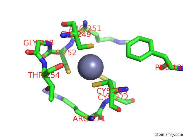
Mono view
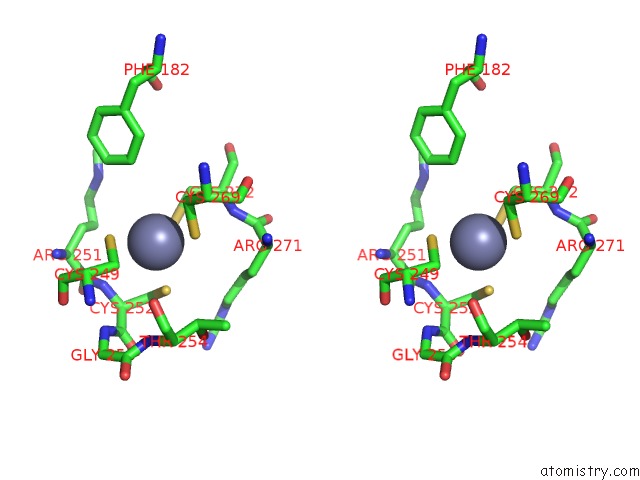
Stereo pair view
Mono view
Stereo pair view
A full contact list of Zinc with other atoms in the Zn binding
site number 1 of Catalytically Inactive (E3Q) Mutm Crosslinked to Oxog:C Containing Dna CC2 within 5.0Å range:
|
Reference:
A.Banerjee,
W.L.Santos,
G.L.Verdine.
Structure of A Dna Glycosylase Searching For Lesions. Science V. 311 1153 2006.
ISSN: ISSN 0036-8075
PubMed: 16497933
DOI: 10.1126/SCIENCE.1120288
Page generated: Wed Aug 20 02:38:14 2025
ISSN: ISSN 0036-8075
PubMed: 16497933
DOI: 10.1126/SCIENCE.1120288
Last articles
Zn in 2YT9Zn in 2YTE
Zn in 2YTD
Zn in 2YTB
Zn in 2YSM
Zn in 2YT5
Zn in 2YTA
Zn in 2YSV
Zn in 2YSP
Zn in 2YSL