Zinc »
PDB 2exf-2fa7 »
2f18 »
Zinc in PDB 2f18: Golgi Alpha-Mannosidase II Complex with (2R,3R,4S)-2-({[(1R)-2- Hydroxy-1-Phenylethyl]Amino}Methyl)Pyrrolidine-3,4-Diol
Enzymatic activity of Golgi Alpha-Mannosidase II Complex with (2R,3R,4S)-2-({[(1R)-2- Hydroxy-1-Phenylethyl]Amino}Methyl)Pyrrolidine-3,4-Diol
All present enzymatic activity of Golgi Alpha-Mannosidase II Complex with (2R,3R,4S)-2-({[(1R)-2- Hydroxy-1-Phenylethyl]Amino}Methyl)Pyrrolidine-3,4-Diol:
3.2.1.114;
3.2.1.114;
Protein crystallography data
The structure of Golgi Alpha-Mannosidase II Complex with (2R,3R,4S)-2-({[(1R)-2- Hydroxy-1-Phenylethyl]Amino}Methyl)Pyrrolidine-3,4-Diol, PDB code: 2f18
was solved by
D.A.Kuntz,
D.R.Rose,
with X-Ray Crystallography technique. A brief refinement statistics is given in the table below:
| Resolution Low / High (Å) | 29.85 / 1.30 |
| Space group | P 21 21 21 |
| Cell size a, b, c (Å), α, β, γ (°) | 68.965, 109.724, 138.911, 90.00, 90.00, 90.00 |
| R / Rfree (%) | 16.8 / 18 |
Zinc Binding Sites:
The binding sites of Zinc atom in the Golgi Alpha-Mannosidase II Complex with (2R,3R,4S)-2-({[(1R)-2- Hydroxy-1-Phenylethyl]Amino}Methyl)Pyrrolidine-3,4-Diol
(pdb code 2f18). This binding sites where shown within
5.0 Angstroms radius around Zinc atom.
In total only one binding site of Zinc was determined in the Golgi Alpha-Mannosidase II Complex with (2R,3R,4S)-2-({[(1R)-2- Hydroxy-1-Phenylethyl]Amino}Methyl)Pyrrolidine-3,4-Diol, PDB code: 2f18:
In total only one binding site of Zinc was determined in the Golgi Alpha-Mannosidase II Complex with (2R,3R,4S)-2-({[(1R)-2- Hydroxy-1-Phenylethyl]Amino}Methyl)Pyrrolidine-3,4-Diol, PDB code: 2f18:
Zinc binding site 1 out of 1 in 2f18
Go back to
Zinc binding site 1 out
of 1 in the Golgi Alpha-Mannosidase II Complex with (2R,3R,4S)-2-({[(1R)-2- Hydroxy-1-Phenylethyl]Amino}Methyl)Pyrrolidine-3,4-Diol
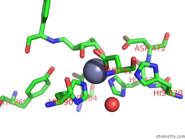
Mono view
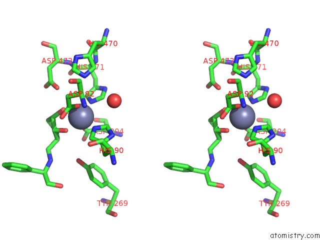
Stereo pair view
Mono view
Stereo pair view
A full contact list of Zinc with other atoms in the Zn binding
site number 1 of Golgi Alpha-Mannosidase II Complex with (2R,3R,4S)-2-({[(1R)-2- Hydroxy-1-Phenylethyl]Amino}Methyl)Pyrrolidine-3,4-Diol within 5.0Å range:
|
Reference:
P.Englebienne,
H.Fiaux,
D.A.Kuntz,
C.R.Corbeil,
S.Gerber-Lemaire,
D.R.Rose,
N.Moitessier.
Evaluation of Docking Programs For Predicting Binding of Golgi Alpha-Mannosidase II Inhibitors: A Comparison with Crystallography. Proteins V. 69 160 2007.
ISSN: ISSN 0887-3585
PubMed: 17557336
DOI: 10.1002/PROT.21479
Page generated: Wed Oct 16 23:36:45 2024
ISSN: ISSN 0887-3585
PubMed: 17557336
DOI: 10.1002/PROT.21479
Last articles
Zn in 9MJ5Zn in 9HNW
Zn in 9G0L
Zn in 9FNE
Zn in 9DZN
Zn in 9E0I
Zn in 9D32
Zn in 9DAK
Zn in 8ZXC
Zn in 8ZUF