Zinc »
PDB 2eex-2elu »
2ejv »
Zinc in PDB 2ejv: Crystal Structure of Threonine 3-Dehydrogenase Complexed with Nad+
Enzymatic activity of Crystal Structure of Threonine 3-Dehydrogenase Complexed with Nad+
All present enzymatic activity of Crystal Structure of Threonine 3-Dehydrogenase Complexed with Nad+:
1.1.1.103;
1.1.1.103;
Protein crystallography data
The structure of Crystal Structure of Threonine 3-Dehydrogenase Complexed with Nad+, PDB code: 2ejv
was solved by
R.Omi,
T.Yao,
M.Goto,
I.Miyahara,
K.Hirotsu,
with X-Ray Crystallography technique. A brief refinement statistics is given in the table below:
| Resolution Low / High (Å) | 19.99 / 2.55 |
| Space group | P 61 2 2 |
| Cell size a, b, c (Å), α, β, γ (°) | 135.758, 135.758, 268.593, 90.00, 90.00, 120.00 |
| R / Rfree (%) | 20.7 / 23.6 |
Zinc Binding Sites:
The binding sites of Zinc atom in the Crystal Structure of Threonine 3-Dehydrogenase Complexed with Nad+
(pdb code 2ejv). This binding sites where shown within
5.0 Angstroms radius around Zinc atom.
In total 4 binding sites of Zinc where determined in the Crystal Structure of Threonine 3-Dehydrogenase Complexed with Nad+, PDB code: 2ejv:
Jump to Zinc binding site number: 1; 2; 3; 4;
In total 4 binding sites of Zinc where determined in the Crystal Structure of Threonine 3-Dehydrogenase Complexed with Nad+, PDB code: 2ejv:
Jump to Zinc binding site number: 1; 2; 3; 4;
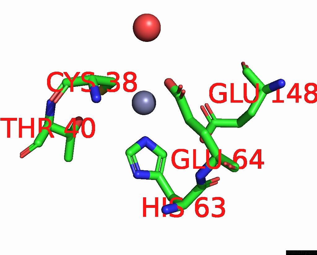
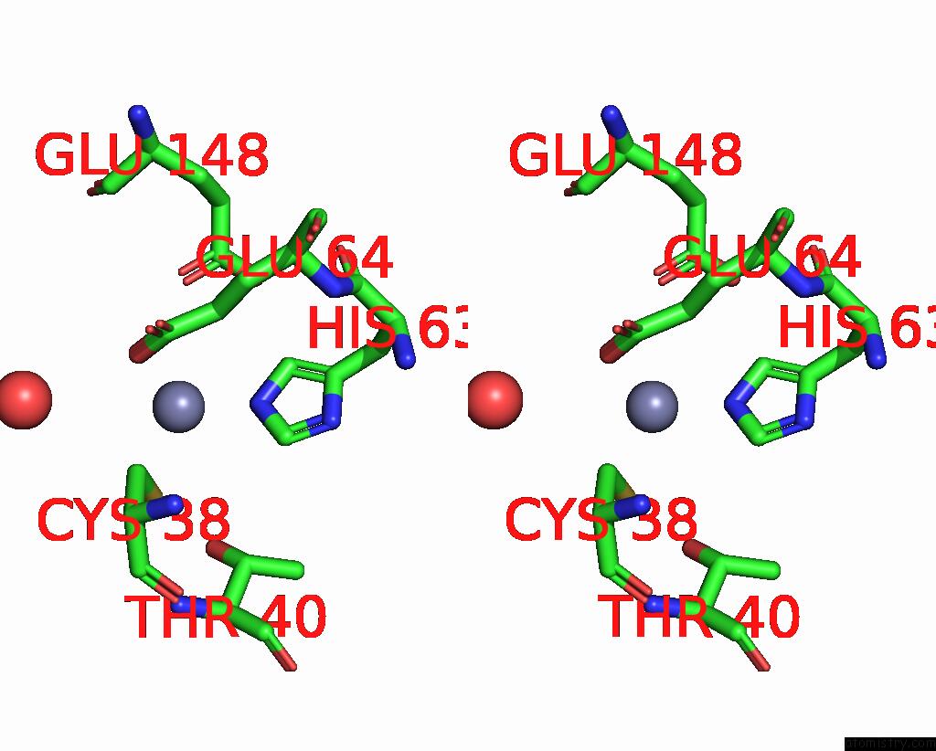
Zinc binding site 1 out of 4 in 2ejv
Go back to
Zinc binding site 1 out
of 4 in the Crystal Structure of Threonine 3-Dehydrogenase Complexed with Nad+
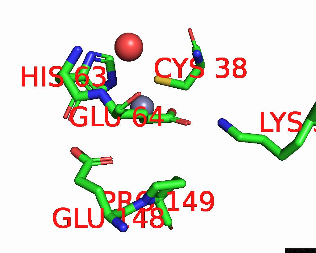
Mono view
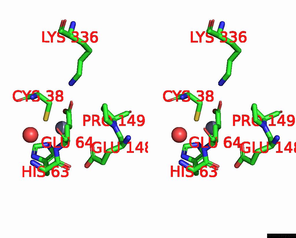
Stereo pair view
Mono view
Stereo pair view
A full contact list of Zinc with other atoms in the Zn binding
site number 1 of Crystal Structure of Threonine 3-Dehydrogenase Complexed with Nad+ within 5.0Å range:
|
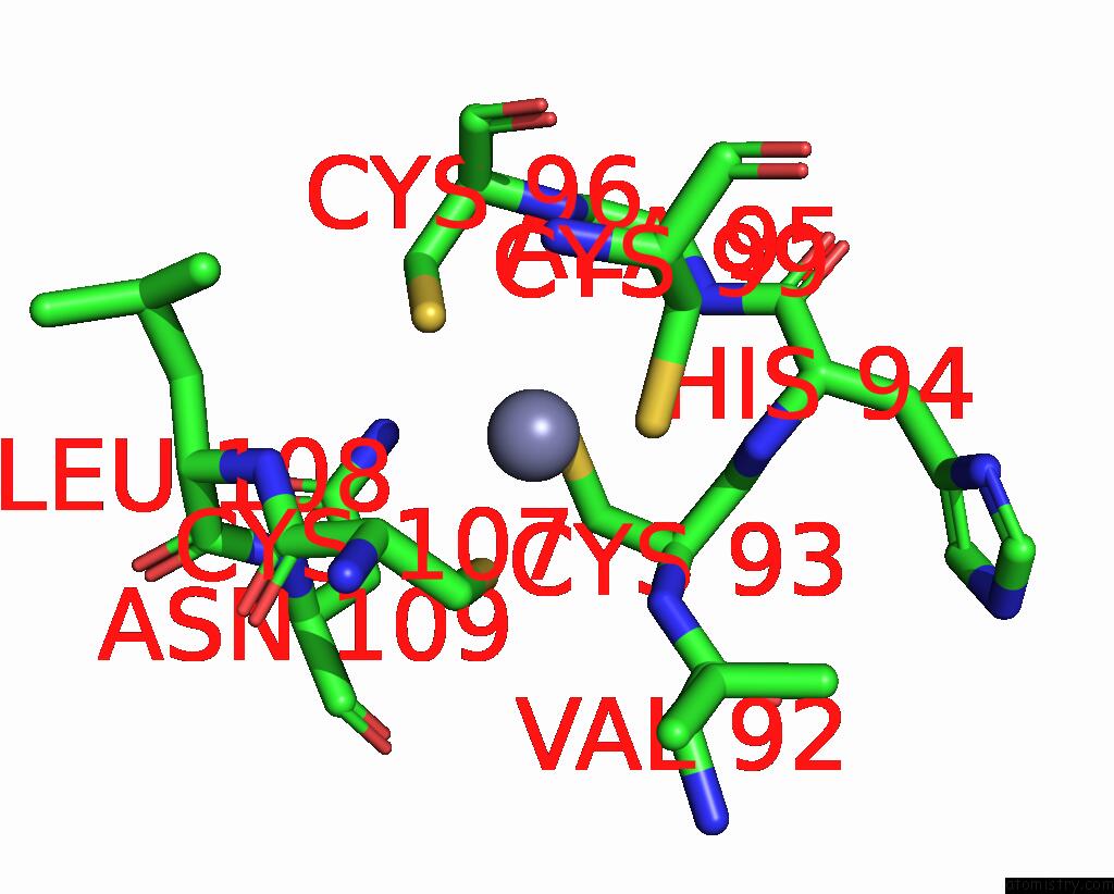
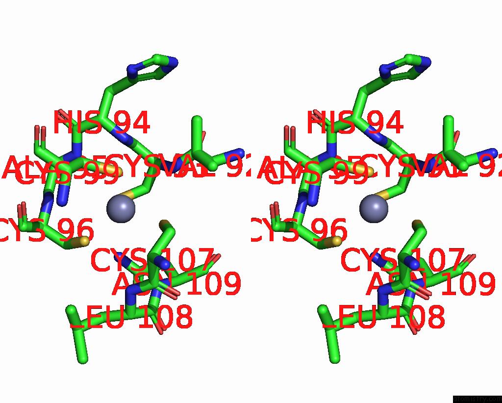
Zinc binding site 2 out of 4 in 2ejv
Go back to
Zinc binding site 2 out
of 4 in the Crystal Structure of Threonine 3-Dehydrogenase Complexed with Nad+
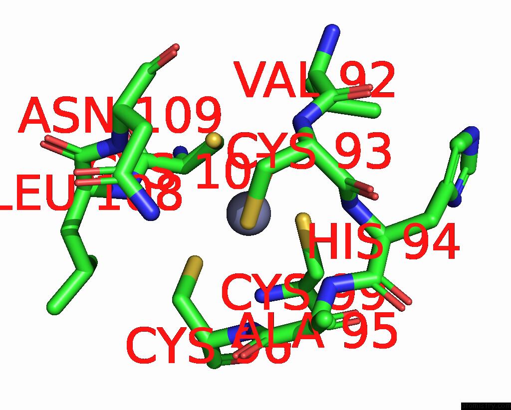
Mono view
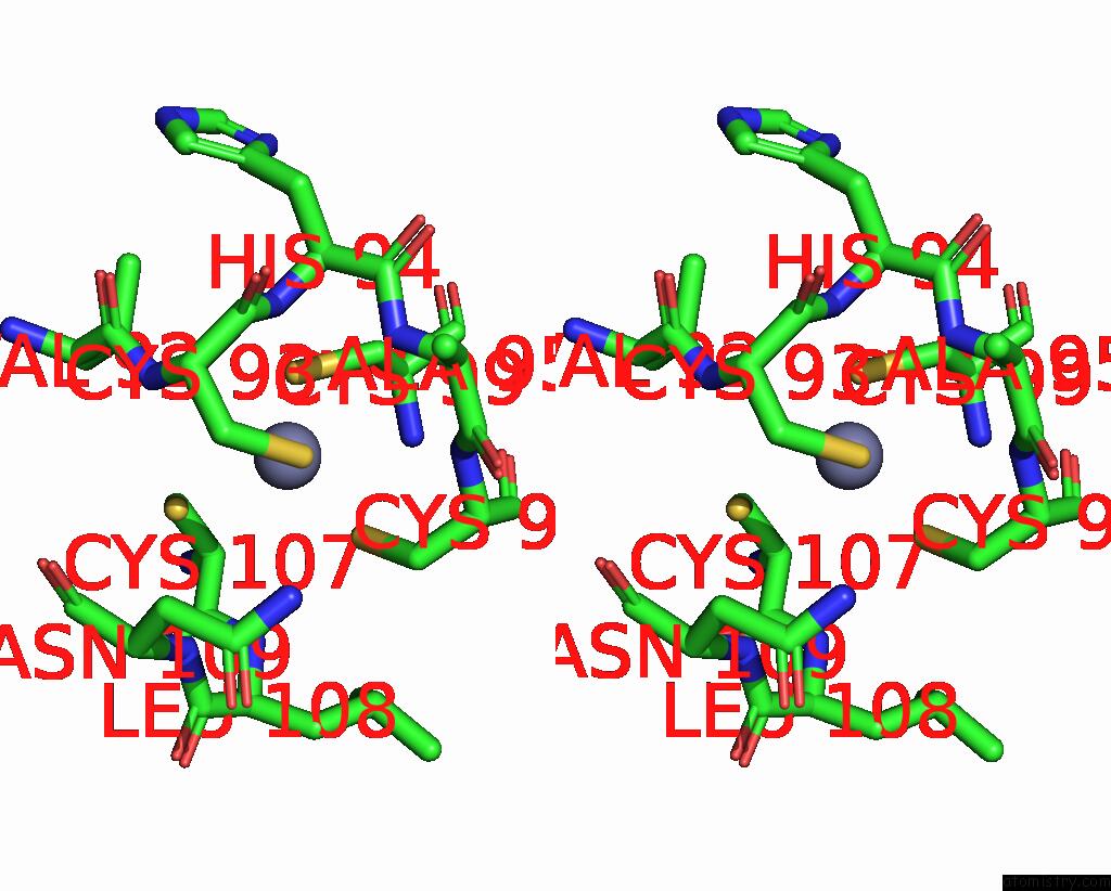
Stereo pair view
Mono view
Stereo pair view
A full contact list of Zinc with other atoms in the Zn binding
site number 2 of Crystal Structure of Threonine 3-Dehydrogenase Complexed with Nad+ within 5.0Å range:
|
Zinc binding site 3 out of 4 in 2ejv
Go back to
Zinc binding site 3 out
of 4 in the Crystal Structure of Threonine 3-Dehydrogenase Complexed with Nad+
Mono view
Stereo pair view
Mono view
Stereo pair view
A full contact list of Zinc with other atoms in the Zn binding
site number 3 of Crystal Structure of Threonine 3-Dehydrogenase Complexed with Nad+ within 5.0Å range:
|
Zinc binding site 4 out of 4 in 2ejv
Go back to
Zinc binding site 4 out
of 4 in the Crystal Structure of Threonine 3-Dehydrogenase Complexed with Nad+
Mono view
Stereo pair view
Mono view
Stereo pair view
A full contact list of Zinc with other atoms in the Zn binding
site number 4 of Crystal Structure of Threonine 3-Dehydrogenase Complexed with Nad+ within 5.0Å range:
|
Reference:
R.Omi,
T.Yao,
M.Goto,
I.Miyahara,
K.Hirotsu.
Crystal Structure of Threonine 3-Dehydrogenase To Be Published.
Page generated: Wed Oct 16 23:12:20 2024
Last articles
Ag in 9GDMAg in 8DYK
Ag in 8GY1
Ag in 8GK2
Ag in 8VW8
Ag in 8VW7
Ag in 8E4D
Ag in 8EG4
Ag in 8E4E
Ag in 7XLW