Zinc »
PDB 2e3x-2eer »
2ebn »
Zinc in PDB 2ebn: Crystal Structure of Endo-Beta-N-Acetylglucosaminidase F1, An Alpha(Slash)Beta-Barrel Enzyme Adapted For A Complex Substrate
Enzymatic activity of Crystal Structure of Endo-Beta-N-Acetylglucosaminidase F1, An Alpha(Slash)Beta-Barrel Enzyme Adapted For A Complex Substrate
All present enzymatic activity of Crystal Structure of Endo-Beta-N-Acetylglucosaminidase F1, An Alpha(Slash)Beta-Barrel Enzyme Adapted For A Complex Substrate:
3.2.1.96;
3.2.1.96;
Protein crystallography data
The structure of Crystal Structure of Endo-Beta-N-Acetylglucosaminidase F1, An Alpha(Slash)Beta-Barrel Enzyme Adapted For A Complex Substrate, PDB code: 2ebn
was solved by
P.Van Roey,
with X-Ray Crystallography technique. A brief refinement statistics is given in the table below:
| Resolution Low / High (Å) | 10.00 / 2.00 |
| Space group | P 61 |
| Cell size a, b, c (Å), α, β, γ (°) | 70.640, 70.640, 103.310, 90.00, 90.00, 120.00 |
| R / Rfree (%) | n/a / n/a |
Zinc Binding Sites:
The binding sites of Zinc atom in the Crystal Structure of Endo-Beta-N-Acetylglucosaminidase F1, An Alpha(Slash)Beta-Barrel Enzyme Adapted For A Complex Substrate
(pdb code 2ebn). This binding sites where shown within
5.0 Angstroms radius around Zinc atom.
In total only one binding site of Zinc was determined in the Crystal Structure of Endo-Beta-N-Acetylglucosaminidase F1, An Alpha(Slash)Beta-Barrel Enzyme Adapted For A Complex Substrate, PDB code: 2ebn:
In total only one binding site of Zinc was determined in the Crystal Structure of Endo-Beta-N-Acetylglucosaminidase F1, An Alpha(Slash)Beta-Barrel Enzyme Adapted For A Complex Substrate, PDB code: 2ebn:
Zinc binding site 1 out of 1 in 2ebn
Go back to
Zinc binding site 1 out
of 1 in the Crystal Structure of Endo-Beta-N-Acetylglucosaminidase F1, An Alpha(Slash)Beta-Barrel Enzyme Adapted For A Complex Substrate
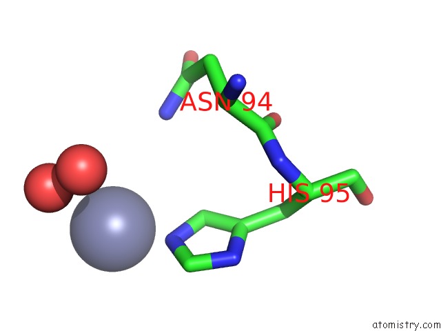
Mono view
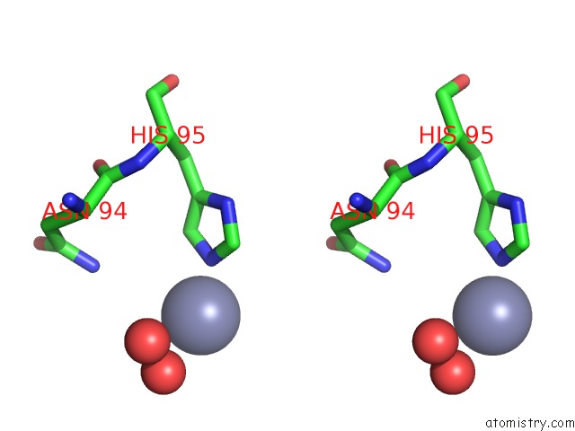
Stereo pair view
Mono view
Stereo pair view
A full contact list of Zinc with other atoms in the Zn binding
site number 1 of Crystal Structure of Endo-Beta-N-Acetylglucosaminidase F1, An Alpha(Slash)Beta-Barrel Enzyme Adapted For A Complex Substrate within 5.0Å range:
|
Reference:
P.Van Roey,
V.Rao,
T.H.Plummer Jr.,
A.L.Tarentino.
Crystal Structure of Endo-Beta-N-Acetylglucosaminidase F1, An Alpha/Beta-Barrel Enzyme Adapted For A Complex Substrate. Biochemistry V. 33 13989 1994.
ISSN: ISSN 0006-2960
PubMed: 7947807
DOI: 10.1021/BI00251A005
Page generated: Wed Oct 16 23:02:31 2024
ISSN: ISSN 0006-2960
PubMed: 7947807
DOI: 10.1021/BI00251A005
Last articles
Zn in 9MJ5Zn in 9HNW
Zn in 9G0L
Zn in 9FNE
Zn in 9DZN
Zn in 9E0I
Zn in 9D32
Zn in 9DAK
Zn in 8ZXC
Zn in 8ZUF