Zinc »
PDB 2e3x-2eer »
2e6l »
Zinc in PDB 2e6l: Structure of Mouse Wrn Exonuclease Domain
Protein crystallography data
The structure of Structure of Mouse Wrn Exonuclease Domain, PDB code: 2e6l
was solved by
Y.Cho,
J.M.Choi,
with X-Ray Crystallography technique. A brief refinement statistics is given in the table below:
| Resolution Low / High (Å) | 29.57 / 2.20 |
| Space group | P 21 21 21 |
| Cell size a, b, c (Å), α, β, γ (°) | 55.047, 59.147, 60.245, 90.00, 90.00, 90.00 |
| R / Rfree (%) | 19.7 / 24.4 |
Zinc Binding Sites:
The binding sites of Zinc atom in the Structure of Mouse Wrn Exonuclease Domain
(pdb code 2e6l). This binding sites where shown within
5.0 Angstroms radius around Zinc atom.
In total 2 binding sites of Zinc where determined in the Structure of Mouse Wrn Exonuclease Domain, PDB code: 2e6l:
Jump to Zinc binding site number: 1; 2;
In total 2 binding sites of Zinc where determined in the Structure of Mouse Wrn Exonuclease Domain, PDB code: 2e6l:
Jump to Zinc binding site number: 1; 2;
Zinc binding site 1 out of 2 in 2e6l
Go back to
Zinc binding site 1 out
of 2 in the Structure of Mouse Wrn Exonuclease Domain
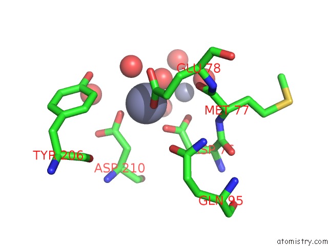
Mono view
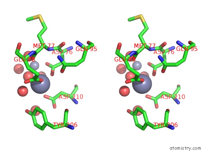
Stereo pair view
Mono view
Stereo pair view
A full contact list of Zinc with other atoms in the Zn binding
site number 1 of Structure of Mouse Wrn Exonuclease Domain within 5.0Å range:
|
Zinc binding site 2 out of 2 in 2e6l
Go back to
Zinc binding site 2 out
of 2 in the Structure of Mouse Wrn Exonuclease Domain
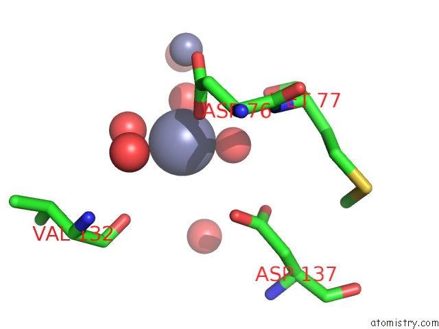
Mono view
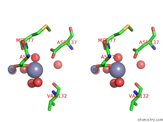
Stereo pair view
Mono view
Stereo pair view
A full contact list of Zinc with other atoms in the Zn binding
site number 2 of Structure of Mouse Wrn Exonuclease Domain within 5.0Å range:
|
Reference:
Y.Cho,
J.M.Choi.
Probing the Roles of Active Site Residues in 3'-5' Exonuclease of Werner Syndrome Protein To Be Published.
Page generated: Wed Aug 20 02:10:49 2025
Last articles
Zn in 2XBLZn in 2XCF
Zn in 2XBQ
Zn in 2XC2
Zn in 2XB3
Zn in 2XAA
Zn in 2XB1
Zn in 2XB4
Zn in 2XAR
Zn in 2XAN