Zinc »
PDB 2ds5-2e2z »
2e25 »
Zinc in PDB 2e25: The Crystal Structure of the T109S Mutant of E. Coli Dihydroorotase Complexed with An Inhibitor 5-Fluoroorotate
Enzymatic activity of The Crystal Structure of the T109S Mutant of E. Coli Dihydroorotase Complexed with An Inhibitor 5-Fluoroorotate
All present enzymatic activity of The Crystal Structure of the T109S Mutant of E. Coli Dihydroorotase Complexed with An Inhibitor 5-Fluoroorotate:
3.5.2.3;
3.5.2.3;
Protein crystallography data
The structure of The Crystal Structure of the T109S Mutant of E. Coli Dihydroorotase Complexed with An Inhibitor 5-Fluoroorotate, PDB code: 2e25
was solved by
M.Lee,
M.J.Maher,
J.M.Guss,
with X-Ray Crystallography technique. A brief refinement statistics is given in the table below:
| Resolution Low / High (Å) | 30.00 / 2.70 |
| Space group | P 41 21 2 |
| Cell size a, b, c (Å), α, β, γ (°) | 72.610, 72.610, 176.120, 90.00, 90.00, 90.00 |
| R / Rfree (%) | 21.8 / 25.7 |
Other elements in 2e25:
The structure of The Crystal Structure of the T109S Mutant of E. Coli Dihydroorotase Complexed with An Inhibitor 5-Fluoroorotate also contains other interesting chemical elements:
| Fluorine | (F) | 1 atom |
Zinc Binding Sites:
The binding sites of Zinc atom in the The Crystal Structure of the T109S Mutant of E. Coli Dihydroorotase Complexed with An Inhibitor 5-Fluoroorotate
(pdb code 2e25). This binding sites where shown within
5.0 Angstroms radius around Zinc atom.
In total 2 binding sites of Zinc where determined in the The Crystal Structure of the T109S Mutant of E. Coli Dihydroorotase Complexed with An Inhibitor 5-Fluoroorotate, PDB code: 2e25:
Jump to Zinc binding site number: 1; 2;
In total 2 binding sites of Zinc where determined in the The Crystal Structure of the T109S Mutant of E. Coli Dihydroorotase Complexed with An Inhibitor 5-Fluoroorotate, PDB code: 2e25:
Jump to Zinc binding site number: 1; 2;
Zinc binding site 1 out of 2 in 2e25
Go back to
Zinc binding site 1 out
of 2 in the The Crystal Structure of the T109S Mutant of E. Coli Dihydroorotase Complexed with An Inhibitor 5-Fluoroorotate
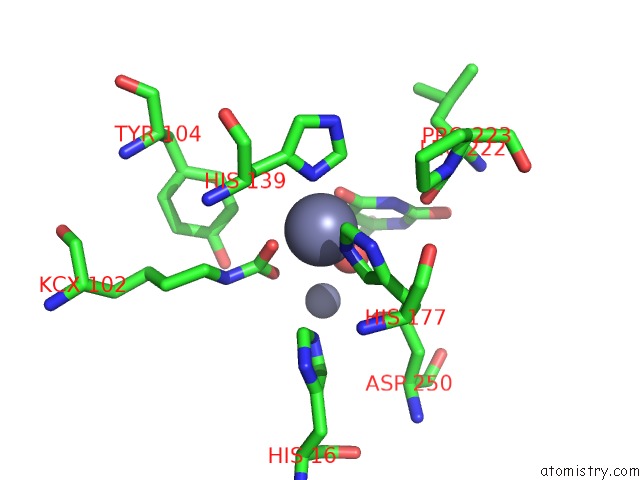
Mono view
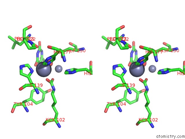
Stereo pair view
Mono view
Stereo pair view
A full contact list of Zinc with other atoms in the Zn binding
site number 1 of The Crystal Structure of the T109S Mutant of E. Coli Dihydroorotase Complexed with An Inhibitor 5-Fluoroorotate within 5.0Å range:
|
Zinc binding site 2 out of 2 in 2e25
Go back to
Zinc binding site 2 out
of 2 in the The Crystal Structure of the T109S Mutant of E. Coli Dihydroorotase Complexed with An Inhibitor 5-Fluoroorotate
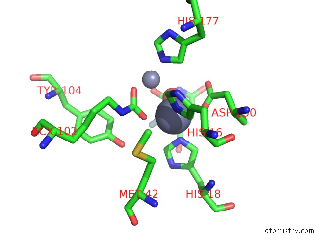
Mono view
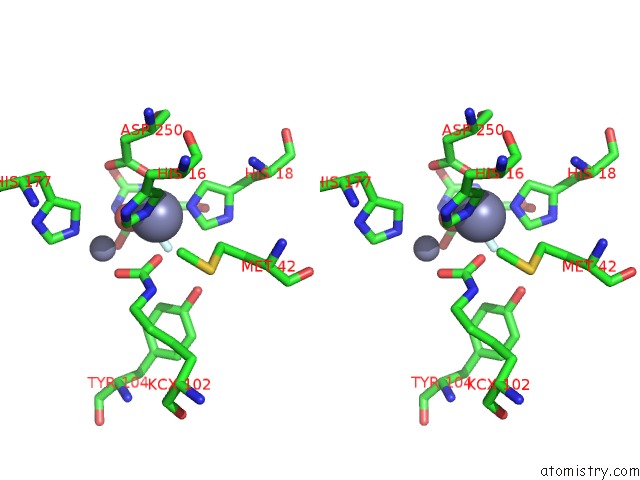
Stereo pair view
Mono view
Stereo pair view
A full contact list of Zinc with other atoms in the Zn binding
site number 2 of The Crystal Structure of the T109S Mutant of E. Coli Dihydroorotase Complexed with An Inhibitor 5-Fluoroorotate within 5.0Å range:
|
Reference:
M.Lee,
M.J.Maher,
J.M.Guss.
Structure of the T109S Mutant of Escherichia Coli Dihydroorotase Complexed with the Inhibitor 5-Fluoroorotate: Catalytic Activity Is Reflected By the Crystal Form Acta Crystallogr.,Sect.F V. 63 154 2007.
ISSN: ESSN 1744-3091
PubMed: 17329804
DOI: 10.1107/S1744309107004009
Page generated: Wed Oct 16 22:56:10 2024
ISSN: ESSN 1744-3091
PubMed: 17329804
DOI: 10.1107/S1744309107004009
Last articles
Zn in 9MJ5Zn in 9HNW
Zn in 9G0L
Zn in 9FNE
Zn in 9DZN
Zn in 9E0I
Zn in 9D32
Zn in 9DAK
Zn in 8ZXC
Zn in 8ZUF