Zinc »
PDB 2ds5-2e2z »
2e1w »
Zinc in PDB 2e1w: Crystal Structure of Adenosine Deaminase Complexed with Potent Inhibitors
Enzymatic activity of Crystal Structure of Adenosine Deaminase Complexed with Potent Inhibitors
All present enzymatic activity of Crystal Structure of Adenosine Deaminase Complexed with Potent Inhibitors:
3.5.4.4;
3.5.4.4;
Protein crystallography data
The structure of Crystal Structure of Adenosine Deaminase Complexed with Potent Inhibitors, PDB code: 2e1w
was solved by
T.Kinoshita,
with X-Ray Crystallography technique. A brief refinement statistics is given in the table below:
| Resolution Low / High (Å) | 29.79 / 2.50 |
| Space group | P 43 21 2 |
| Cell size a, b, c (Å), α, β, γ (°) | 78.410, 78.410, 137.460, 90.00, 90.00, 90.00 |
| R / Rfree (%) | n/a / n/a |
Zinc Binding Sites:
The binding sites of Zinc atom in the Crystal Structure of Adenosine Deaminase Complexed with Potent Inhibitors
(pdb code 2e1w). This binding sites where shown within
5.0 Angstroms radius around Zinc atom.
In total only one binding site of Zinc was determined in the Crystal Structure of Adenosine Deaminase Complexed with Potent Inhibitors, PDB code: 2e1w:
In total only one binding site of Zinc was determined in the Crystal Structure of Adenosine Deaminase Complexed with Potent Inhibitors, PDB code: 2e1w:
Zinc binding site 1 out of 1 in 2e1w
Go back to
Zinc binding site 1 out
of 1 in the Crystal Structure of Adenosine Deaminase Complexed with Potent Inhibitors
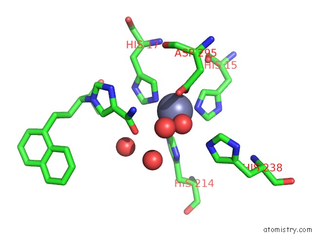
Mono view
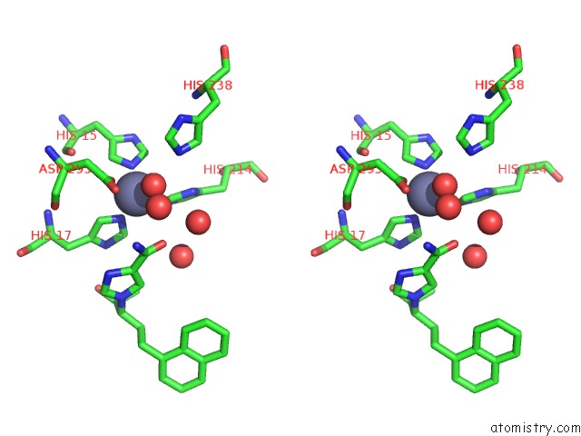
Stereo pair view
Mono view
Stereo pair view
A full contact list of Zinc with other atoms in the Zn binding
site number 1 of Crystal Structure of Adenosine Deaminase Complexed with Potent Inhibitors within 5.0Å range:
|
Reference:
T.Terasaka,
H.Okumura,
K.Tsuji,
T.Kato,
I.Nakanishi,
T.Kinoshita,
Y.Kato,
M.Kuno,
N.Seki,
Y.Naoe,
T.Inoue,
K.Tanaka,
K.Nakamura.
Structure-Based Design and Synthesis of Non-Nucleoside, Potent, and Orally Bioavailable Adenosine Deaminase Inhibitors J.Med.Chem. V. 47 2728 2004.
ISSN: ISSN 0022-2623
PubMed: 15139750
DOI: 10.1021/JM0499559
Page generated: Wed Oct 16 22:55:53 2024
ISSN: ISSN 0022-2623
PubMed: 15139750
DOI: 10.1021/JM0499559
Last articles
Zn in 9MJ5Zn in 9HNW
Zn in 9G0L
Zn in 9FNE
Zn in 9DZN
Zn in 9E0I
Zn in 9D32
Zn in 9DAK
Zn in 8ZXC
Zn in 8ZUF