Zinc »
PDB 2db6-2drp »
2dqm »
Zinc in PDB 2dqm: Crystal Structure of Aminopeptidase N Complexed with Bestatin
Enzymatic activity of Crystal Structure of Aminopeptidase N Complexed with Bestatin
All present enzymatic activity of Crystal Structure of Aminopeptidase N Complexed with Bestatin:
3.4.11.2;
3.4.11.2;
Protein crystallography data
The structure of Crystal Structure of Aminopeptidase N Complexed with Bestatin, PDB code: 2dqm
was solved by
Y.Onohara,
Y.Nakajima,
K.Ito,
T.Yoshimoto,
with X-Ray Crystallography technique. A brief refinement statistics is given in the table below:
| Resolution Low / High (Å) | 20.00 / 1.60 |
| Space group | P 31 2 1 |
| Cell size a, b, c (Å), α, β, γ (°) | 120.417, 120.417, 170.859, 90.00, 90.00, 120.00 |
| R / Rfree (%) | 18.2 / 19.4 |
Zinc Binding Sites:
The binding sites of Zinc atom in the Crystal Structure of Aminopeptidase N Complexed with Bestatin
(pdb code 2dqm). This binding sites where shown within
5.0 Angstroms radius around Zinc atom.
In total only one binding site of Zinc was determined in the Crystal Structure of Aminopeptidase N Complexed with Bestatin, PDB code: 2dqm:
In total only one binding site of Zinc was determined in the Crystal Structure of Aminopeptidase N Complexed with Bestatin, PDB code: 2dqm:
Zinc binding site 1 out of 1 in 2dqm
Go back to
Zinc binding site 1 out
of 1 in the Crystal Structure of Aminopeptidase N Complexed with Bestatin
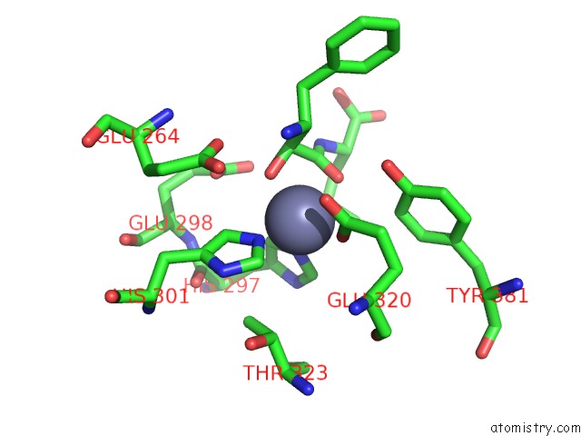
Mono view
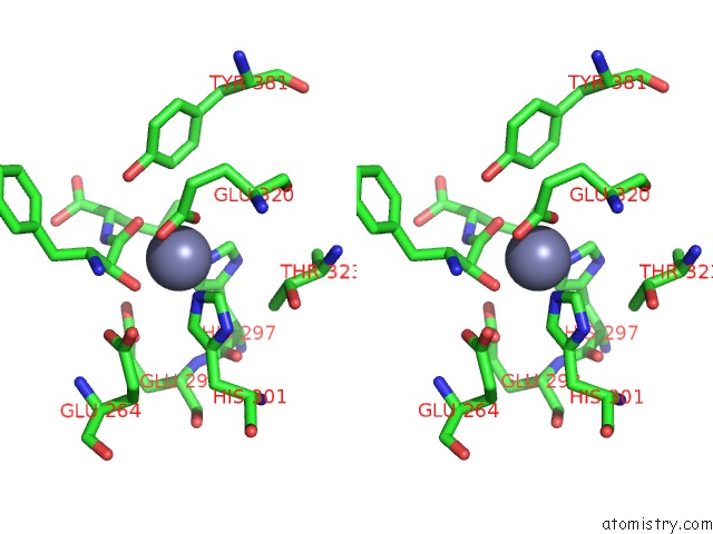
Stereo pair view
Mono view
Stereo pair view
A full contact list of Zinc with other atoms in the Zn binding
site number 1 of Crystal Structure of Aminopeptidase N Complexed with Bestatin within 5.0Å range:
|
Reference:
K.Ito,
Y.Nakajima,
Y.Onohara,
M.Takeo,
K.Nakashima,
F.Matsubara,
T.Ito,
T.Yoshimoto.
Aminopeptidase N (Proteobacteria Alanyl Aminopeptidase) From Escherichia Coli: Crystal Structure and Conformational Change of the Methionine 260 Residue Involved in Substrate Recognition J.Biol.Chem. V. 281 33664 2006.
ISSN: ISSN 0021-9258
PubMed: 16885166
DOI: 10.1074/JBC.M605203200
Page generated: Wed Oct 16 22:50:33 2024
ISSN: ISSN 0021-9258
PubMed: 16885166
DOI: 10.1074/JBC.M605203200
Last articles
Zn in 9MJ5Zn in 9HNW
Zn in 9G0L
Zn in 9FNE
Zn in 9DZN
Zn in 9E0I
Zn in 9D32
Zn in 9DAK
Zn in 8ZXC
Zn in 8ZUF