Zinc »
PDB 2c7a-2cij »
2ci6 »
Zinc in PDB 2ci6: Crystal Structure of Dimethylarginine Dimethylaminohydrolase I Bound with Zinc Low pH
Enzymatic activity of Crystal Structure of Dimethylarginine Dimethylaminohydrolase I Bound with Zinc Low pH
All present enzymatic activity of Crystal Structure of Dimethylarginine Dimethylaminohydrolase I Bound with Zinc Low pH:
3.5.3.18;
3.5.3.18;
Protein crystallography data
The structure of Crystal Structure of Dimethylarginine Dimethylaminohydrolase I Bound with Zinc Low pH, PDB code: 2ci6
was solved by
D.Frey,
O.Braun,
C.Briand,
M.Vasak,
M.G.Grutter,
with X-Ray Crystallography technique. A brief refinement statistics is given in the table below:
| Resolution Low / High (Å) | 39.08 / 2.0 |
| Space group | P 21 21 21 |
| Cell size a, b, c (Å), α, β, γ (°) | 44.500, 76.670, 81.670, 90.00, 90.00, 90.00 |
| R / Rfree (%) | 23.8 / 26.3 |
Zinc Binding Sites:
The binding sites of Zinc atom in the Crystal Structure of Dimethylarginine Dimethylaminohydrolase I Bound with Zinc Low pH
(pdb code 2ci6). This binding sites where shown within
5.0 Angstroms radius around Zinc atom.
In total only one binding site of Zinc was determined in the Crystal Structure of Dimethylarginine Dimethylaminohydrolase I Bound with Zinc Low pH, PDB code: 2ci6:
In total only one binding site of Zinc was determined in the Crystal Structure of Dimethylarginine Dimethylaminohydrolase I Bound with Zinc Low pH, PDB code: 2ci6:
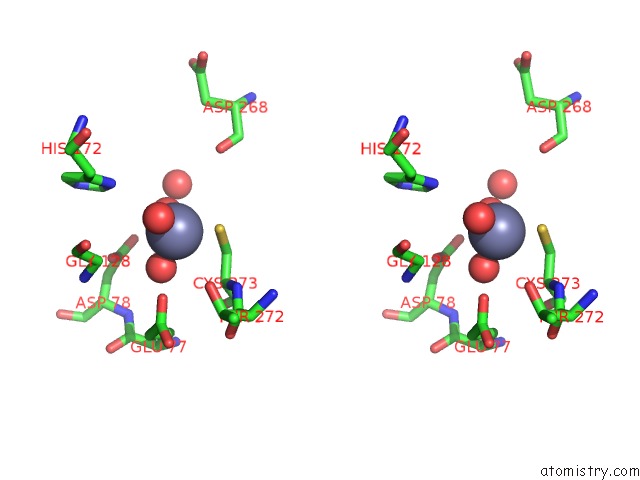
Zinc binding site 1 out of 1 in 2ci6
Go back to
Zinc binding site 1 out
of 1 in the Crystal Structure of Dimethylarginine Dimethylaminohydrolase I Bound with Zinc Low pH

Mono view
Stereo pair view
Mono view
Stereo pair view
A full contact list of Zinc with other atoms in the Zn binding
site number 1 of Crystal Structure of Dimethylarginine Dimethylaminohydrolase I Bound with Zinc Low pH within 5.0Å range:
|
Reference:
D.Frey,
O.Braun,
C.Briand,
M.Vasak,
M.G.Grutter.
Structure of the Mammalian Nos Regulator Dimethylarginine Dimethylaminohydrolase: A Basis For the Design of Specific Inbitors Structure V. 14 901 2006.
ISSN: ISSN 0969-2126
PubMed: 16698551
DOI: 10.1016/J.STR.2006.03.006
Page generated: Wed Aug 20 01:42:54 2025
ISSN: ISSN 0969-2126
PubMed: 16698551
DOI: 10.1016/J.STR.2006.03.006
Last articles
Zn in 2W8CZn in 2W5W
Zn in 2W5X
Zn in 2W5Z
Zn in 2W5Y
Zn in 2W4L
Zn in 2W5V
Zn in 2W57
Zn in 2W3N
Zn in 2W44