Zinc »
PDB 2bnn-2c6w »
2bz1 »
Zinc in PDB 2bz1: Crystal Structure of Apo E. Coli Gtp Cyclohydrolase II
Enzymatic activity of Crystal Structure of Apo E. Coli Gtp Cyclohydrolase II
All present enzymatic activity of Crystal Structure of Apo E. Coli Gtp Cyclohydrolase II:
3.5.4.25;
3.5.4.25;
Protein crystallography data
The structure of Crystal Structure of Apo E. Coli Gtp Cyclohydrolase II, PDB code: 2bz1
was solved by
J.Ren,
M.Kotaka,
M.Lockyer,
H.K.Lamb,
A.R.Hawkins,
D.K.Stammers,
with X-Ray Crystallography technique. A brief refinement statistics is given in the table below:
| Resolution Low / High (Å) | 28.57 / 1.54 |
| Space group | P 65 2 2 |
| Cell size a, b, c (Å), α, β, γ (°) | 71.920, 71.920, 128.630, 90.00, 90.00, 120.00 |
| R / Rfree (%) | 19.3 / 21.2 |
Zinc Binding Sites:
The binding sites of Zinc atom in the Crystal Structure of Apo E. Coli Gtp Cyclohydrolase II
(pdb code 2bz1). This binding sites where shown within
5.0 Angstroms radius around Zinc atom.
In total only one binding site of Zinc was determined in the Crystal Structure of Apo E. Coli Gtp Cyclohydrolase II, PDB code: 2bz1:
In total only one binding site of Zinc was determined in the Crystal Structure of Apo E. Coli Gtp Cyclohydrolase II, PDB code: 2bz1:
Zinc binding site 1 out of 1 in 2bz1
Go back to
Zinc binding site 1 out
of 1 in the Crystal Structure of Apo E. Coli Gtp Cyclohydrolase II
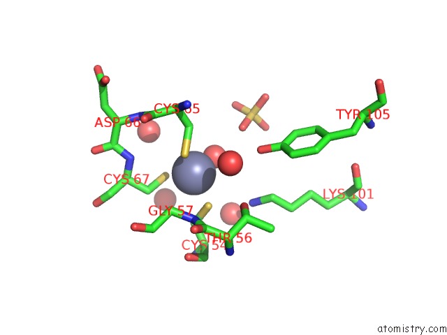
Mono view
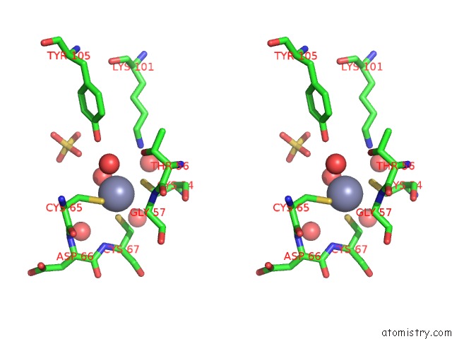
Stereo pair view
Mono view
Stereo pair view
A full contact list of Zinc with other atoms in the Zn binding
site number 1 of Crystal Structure of Apo E. Coli Gtp Cyclohydrolase II within 5.0Å range:
|
Reference:
J.Ren,
M.Kotaka,
M.Lockyer,
H.K.Lamb,
A.R.Hawkins,
D.K.Stammers.
Gtp Cyclohydrolase II Structure and Mechanism. J.Biol.Chem. V. 280 36912 2005.
ISSN: ISSN 0021-9258
PubMed: 16115872
DOI: 10.1074/JBC.M507725200
Page generated: Wed Oct 16 22:08:44 2024
ISSN: ISSN 0021-9258
PubMed: 16115872
DOI: 10.1074/JBC.M507725200
Last articles
As in 2XZ1As in 2Y0O
As in 2XQ9
As in 2XOE
As in 2XOD
As in 2XQ4
As in 2XQ5
As in 2VQG
As in 2XNQ
As in 2XFV