Zinc »
PDB 2bnn-2c6w »
2bnn »
Zinc in PDB 2bnn: The Structure of Hydroxypropylphosphonic Acid Epoxidase From S. Wedmorenis in Complex with Fosfomycin
Protein crystallography data
The structure of The Structure of Hydroxypropylphosphonic Acid Epoxidase From S. Wedmorenis in Complex with Fosfomycin, PDB code: 2bnn
was solved by
K.Mcluskey,
S.Cameron,
W.N.Hunter,
with X-Ray Crystallography technique. A brief refinement statistics is given in the table below:
| Resolution Low / High (Å) | 30.00 / 2.50 |
| Space group | P 65 2 2 |
| Cell size a, b, c (Å), α, β, γ (°) | 85.602, 85.602, 217.989, 90.00, 90.00, 120.00 |
| R / Rfree (%) | 18.4 / 27 |
Zinc Binding Sites:
The binding sites of Zinc atom in the The Structure of Hydroxypropylphosphonic Acid Epoxidase From S. Wedmorenis in Complex with Fosfomycin
(pdb code 2bnn). This binding sites where shown within
5.0 Angstroms radius around Zinc atom.
In total 2 binding sites of Zinc where determined in the The Structure of Hydroxypropylphosphonic Acid Epoxidase From S. Wedmorenis in Complex with Fosfomycin, PDB code: 2bnn:
Jump to Zinc binding site number: 1; 2;
In total 2 binding sites of Zinc where determined in the The Structure of Hydroxypropylphosphonic Acid Epoxidase From S. Wedmorenis in Complex with Fosfomycin, PDB code: 2bnn:
Jump to Zinc binding site number: 1; 2;
Zinc binding site 1 out of 2 in 2bnn
Go back to
Zinc binding site 1 out
of 2 in the The Structure of Hydroxypropylphosphonic Acid Epoxidase From S. Wedmorenis in Complex with Fosfomycin
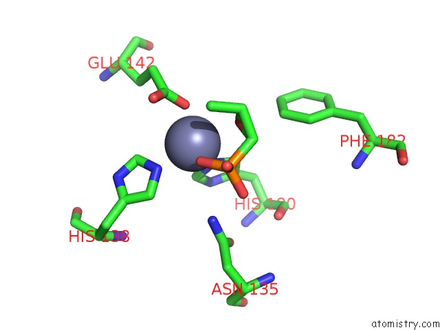
Mono view
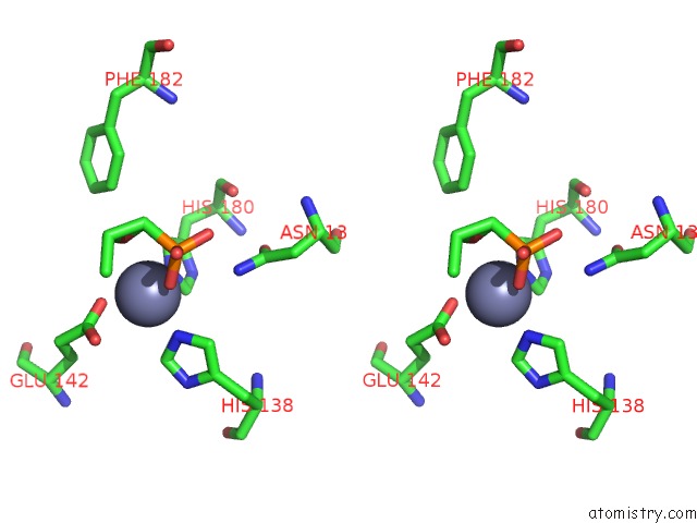
Stereo pair view
Mono view
Stereo pair view
A full contact list of Zinc with other atoms in the Zn binding
site number 1 of The Structure of Hydroxypropylphosphonic Acid Epoxidase From S. Wedmorenis in Complex with Fosfomycin within 5.0Å range:
|
Zinc binding site 2 out of 2 in 2bnn
Go back to
Zinc binding site 2 out
of 2 in the The Structure of Hydroxypropylphosphonic Acid Epoxidase From S. Wedmorenis in Complex with Fosfomycin
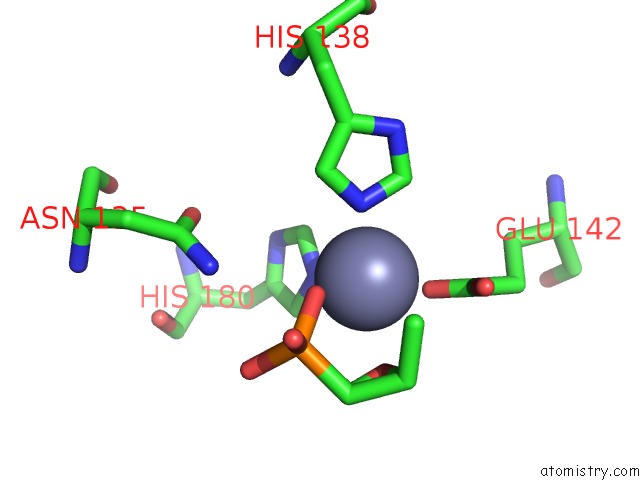
Mono view
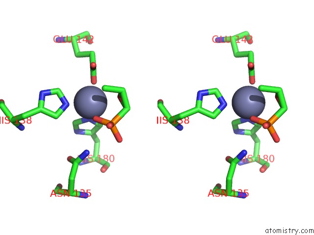
Stereo pair view
Mono view
Stereo pair view
A full contact list of Zinc with other atoms in the Zn binding
site number 2 of The Structure of Hydroxypropylphosphonic Acid Epoxidase From S. Wedmorenis in Complex with Fosfomycin within 5.0Å range:
|
Reference:
K.Mcluskey,
S.Cameron,
F.Hammerschmidt,
W.N.Hunter.
Structure and Reactivity of Hydroxypropylphosphonic Acid Epoxidase in Fosfomycin Biosynthesis By A Cation- and Flavin-Dependent Mechanism. Proc.Natl.Acad.Sci.Usa V. 102 14221 2005.
ISSN: ISSN 0027-8424
PubMed: 16186494
DOI: 10.1073/PNAS.0504314102
Page generated: Wed Oct 16 22:04:19 2024
ISSN: ISSN 0027-8424
PubMed: 16186494
DOI: 10.1073/PNAS.0504314102
Last articles
Zn in 9MJ5Zn in 9HNW
Zn in 9G0L
Zn in 9FNE
Zn in 9DZN
Zn in 9E0I
Zn in 9D32
Zn in 9DAK
Zn in 8ZXC
Zn in 8ZUF