Zinc »
PDB 2aqs-2b83 »
2b0z »
Zinc in PDB 2b0z: Crystal Structure of the Protein-Protein Complex Between F82I Cytochrome C and Cytochrome C Peroxidase
Enzymatic activity of Crystal Structure of the Protein-Protein Complex Between F82I Cytochrome C and Cytochrome C Peroxidase
All present enzymatic activity of Crystal Structure of the Protein-Protein Complex Between F82I Cytochrome C and Cytochrome C Peroxidase:
1.11.1.5;
1.11.1.5;
Protein crystallography data
The structure of Crystal Structure of the Protein-Protein Complex Between F82I Cytochrome C and Cytochrome C Peroxidase, PDB code: 2b0z
was solved by
S.A.Kang,
B.R.Crane,
with X-Ray Crystallography technique. A brief refinement statistics is given in the table below:
| Resolution Low / High (Å) | 20.00 / 2.70 |
| Space group | P 21 21 21 |
| Cell size a, b, c (Å), α, β, γ (°) | 43.802, 51.989, 183.669, 90.00, 90.00, 90.00 |
| R / Rfree (%) | 26.2 / 28.9 |
Other elements in 2b0z:
The structure of Crystal Structure of the Protein-Protein Complex Between F82I Cytochrome C and Cytochrome C Peroxidase also contains other interesting chemical elements:
| Iron | (Fe) | 1 atom |
Zinc Binding Sites:
The binding sites of Zinc atom in the Crystal Structure of the Protein-Protein Complex Between F82I Cytochrome C and Cytochrome C Peroxidase
(pdb code 2b0z). This binding sites where shown within
5.0 Angstroms radius around Zinc atom.
In total only one binding site of Zinc was determined in the Crystal Structure of the Protein-Protein Complex Between F82I Cytochrome C and Cytochrome C Peroxidase, PDB code: 2b0z:
In total only one binding site of Zinc was determined in the Crystal Structure of the Protein-Protein Complex Between F82I Cytochrome C and Cytochrome C Peroxidase, PDB code: 2b0z:
Zinc binding site 1 out of 1 in 2b0z
Go back to
Zinc binding site 1 out
of 1 in the Crystal Structure of the Protein-Protein Complex Between F82I Cytochrome C and Cytochrome C Peroxidase
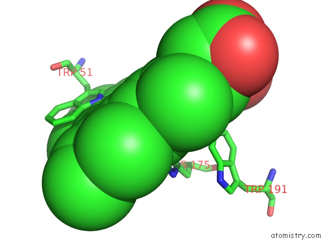
Mono view
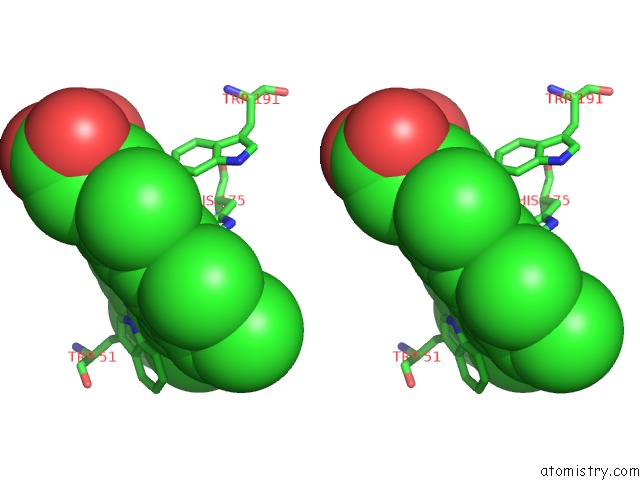
Stereo pair view
Mono view
Stereo pair view
A full contact list of Zinc with other atoms in the Zn binding
site number 1 of Crystal Structure of the Protein-Protein Complex Between F82I Cytochrome C and Cytochrome C Peroxidase within 5.0Å range:
|
Reference:
S.A.Kang,
B.R.Crane.
Effects of Interface Mutations on Association Modes and Electron-Transfer Rates Between Proteins Proc.Natl.Acad.Sci.Usa V. 102 15465 2005.
ISSN: ISSN 0027-8424
PubMed: 16227441
DOI: 10.1073/PNAS.0505176102
Page generated: Wed Oct 16 21:52:33 2024
ISSN: ISSN 0027-8424
PubMed: 16227441
DOI: 10.1073/PNAS.0505176102
Last articles
Zn in 9MJ5Zn in 9HNW
Zn in 9G0L
Zn in 9FNE
Zn in 9DZN
Zn in 9E0I
Zn in 9D32
Zn in 9DAK
Zn in 8ZXC
Zn in 8ZUF