Zinc »
PDB 2aqs-2b83 »
2ata »
Zinc in PDB 2ata: Structural Basis of Dna Recognition By P53 Tetramers (Complex II)
Protein crystallography data
The structure of Structural Basis of Dna Recognition By P53 Tetramers (Complex II), PDB code: 2ata
was solved by
M.Kitayner,
H.Rozenberg,
N.Kessler,
D.Rabinovich,
Z.Shakked,
with X-Ray Crystallography technique. A brief refinement statistics is given in the table below:
| Resolution Low / High (Å) | 43.00 / 2.20 |
| Space group | P 1 |
| Cell size a, b, c (Å), α, β, γ (°) | 54.649, 57.998, 77.983, 83.35, 87.55, 73.50 |
| R / Rfree (%) | 14.5 / 21.5 |
Zinc Binding Sites:
The binding sites of Zinc atom in the Structural Basis of Dna Recognition By P53 Tetramers (Complex II)
(pdb code 2ata). This binding sites where shown within
5.0 Angstroms radius around Zinc atom.
In total 4 binding sites of Zinc where determined in the Structural Basis of Dna Recognition By P53 Tetramers (Complex II), PDB code: 2ata:
Jump to Zinc binding site number: 1; 2; 3; 4;
In total 4 binding sites of Zinc where determined in the Structural Basis of Dna Recognition By P53 Tetramers (Complex II), PDB code: 2ata:
Jump to Zinc binding site number: 1; 2; 3; 4;
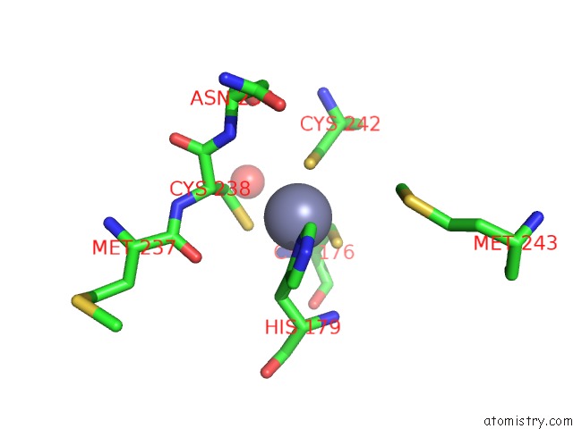
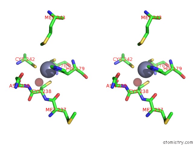
Zinc binding site 1 out of 4 in 2ata
Go back to
Zinc binding site 1 out
of 4 in the Structural Basis of Dna Recognition By P53 Tetramers (Complex II)
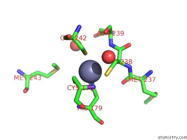
Mono view
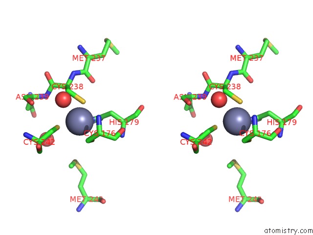
Stereo pair view
Mono view
Stereo pair view
A full contact list of Zinc with other atoms in the Zn binding
site number 1 of Structural Basis of Dna Recognition By P53 Tetramers (Complex II) within 5.0Å range:
|
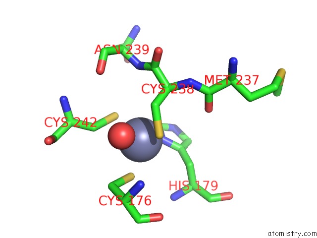
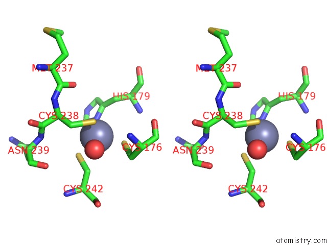
Zinc binding site 2 out of 4 in 2ata
Go back to
Zinc binding site 2 out
of 4 in the Structural Basis of Dna Recognition By P53 Tetramers (Complex II)
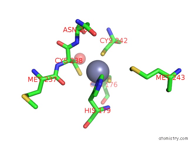
Mono view
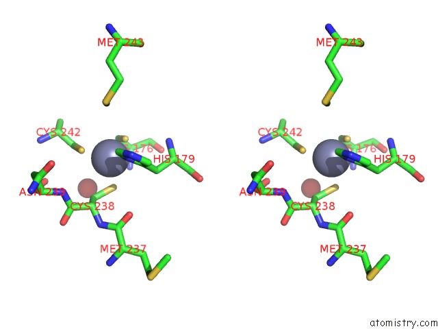
Stereo pair view
Mono view
Stereo pair view
A full contact list of Zinc with other atoms in the Zn binding
site number 2 of Structural Basis of Dna Recognition By P53 Tetramers (Complex II) within 5.0Å range:
|
Zinc binding site 3 out of 4 in 2ata
Go back to
Zinc binding site 3 out
of 4 in the Structural Basis of Dna Recognition By P53 Tetramers (Complex II)
Mono view
Stereo pair view
Mono view
Stereo pair view
A full contact list of Zinc with other atoms in the Zn binding
site number 3 of Structural Basis of Dna Recognition By P53 Tetramers (Complex II) within 5.0Å range:
|
Zinc binding site 4 out of 4 in 2ata
Go back to
Zinc binding site 4 out
of 4 in the Structural Basis of Dna Recognition By P53 Tetramers (Complex II)
Mono view
Stereo pair view
Mono view
Stereo pair view
A full contact list of Zinc with other atoms in the Zn binding
site number 4 of Structural Basis of Dna Recognition By P53 Tetramers (Complex II) within 5.0Å range:
|
Reference:
M.Kitayner,
H.Rozenberg,
N.Kessler,
D.Rabinovich,
L.Shaulov,
T.E.Haran,
Z.Shakked.
Structural Basis of Dna Recognition By P53 Tetramers Mol.Cell V. 22 741 2006.
ISSN: ISSN 1097-2765
PubMed: 16793544
DOI: 10.1016/J.MOLCEL.2006.05.015
Page generated: Wed Oct 16 21:49:11 2024
ISSN: ISSN 1097-2765
PubMed: 16793544
DOI: 10.1016/J.MOLCEL.2006.05.015
Last articles
Zn in 9MJ5Zn in 9HNW
Zn in 9G0L
Zn in 9FNE
Zn in 9DZN
Zn in 9E0I
Zn in 9D32
Zn in 9DAK
Zn in 8ZXC
Zn in 8ZUF