Zinc »
PDB 2afo-2aqr »
2akf »
Zinc in PDB 2akf: Crystal Structure of the Coiled-Coil Domain of Coronin 1
Protein crystallography data
The structure of Crystal Structure of the Coiled-Coil Domain of Coronin 1, PDB code: 2akf
was solved by
R.A.Kammerer,
D.Kostrewa,
P.Progias,
S.Honnappa,
D.Avila,
A.Lustig,
F.K.Winkler,
J.Pieters,
M.O.Steinmetz,
with X-Ray Crystallography technique. A brief refinement statistics is given in the table below:
| Resolution Low / High (Å) | 20.00 / 1.20 |
| Space group | P 1 |
| Cell size a, b, c (Å), α, β, γ (°) | 23.587, 23.569, 46.402, 92.51, 96.85, 119.63 |
| R / Rfree (%) | 16 / 19.6 |
Zinc Binding Sites:
The binding sites of Zinc atom in the Crystal Structure of the Coiled-Coil Domain of Coronin 1
(pdb code 2akf). This binding sites where shown within
5.0 Angstroms radius around Zinc atom.
In total 7 binding sites of Zinc where determined in the Crystal Structure of the Coiled-Coil Domain of Coronin 1, PDB code: 2akf:
Jump to Zinc binding site number: 1; 2; 3; 4; 5; 6; 7;
In total 7 binding sites of Zinc where determined in the Crystal Structure of the Coiled-Coil Domain of Coronin 1, PDB code: 2akf:
Jump to Zinc binding site number: 1; 2; 3; 4; 5; 6; 7;
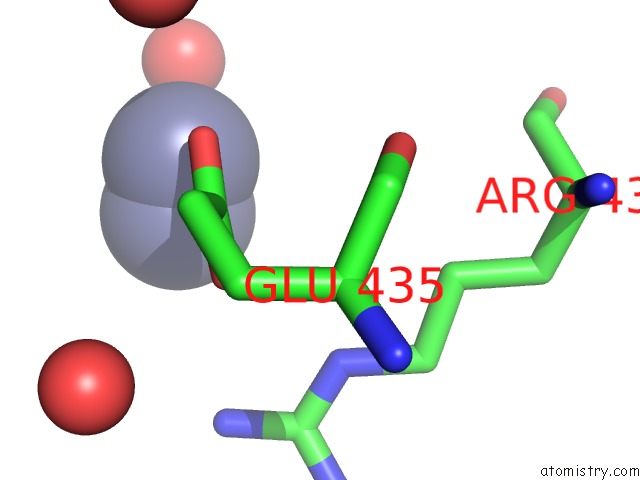
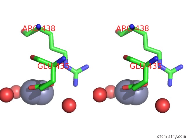
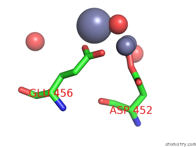
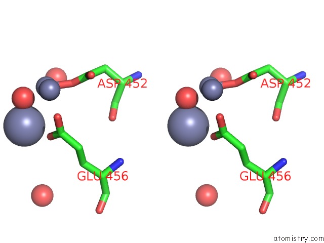
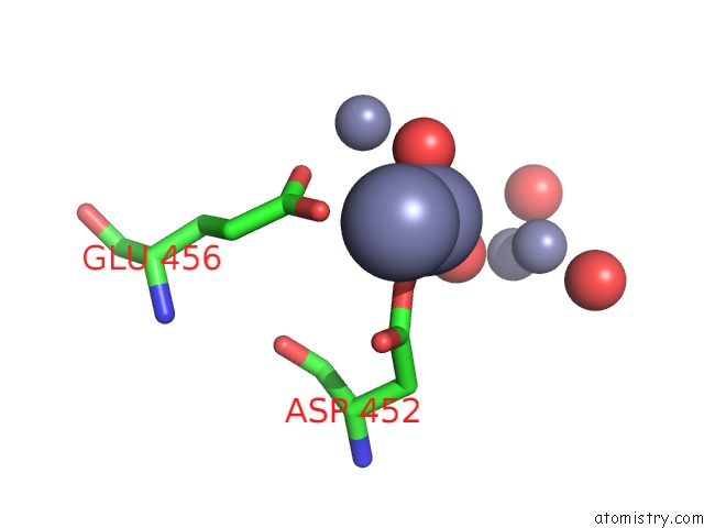
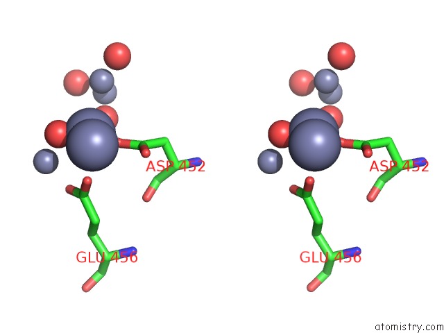
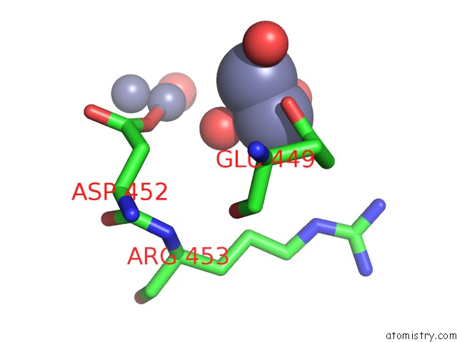
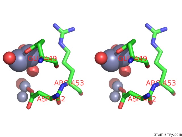
Zinc binding site 1 out of 7 in 2akf
Go back to
Zinc binding site 1 out
of 7 in the Crystal Structure of the Coiled-Coil Domain of Coronin 1
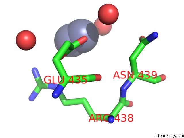
Mono view
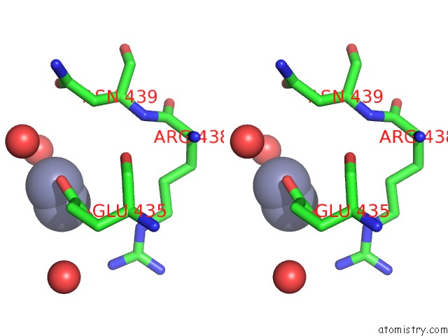
Stereo pair view
Mono view
Stereo pair view
A full contact list of Zinc with other atoms in the Zn binding
site number 1 of Crystal Structure of the Coiled-Coil Domain of Coronin 1 within 5.0Å range:
|
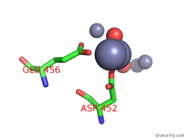
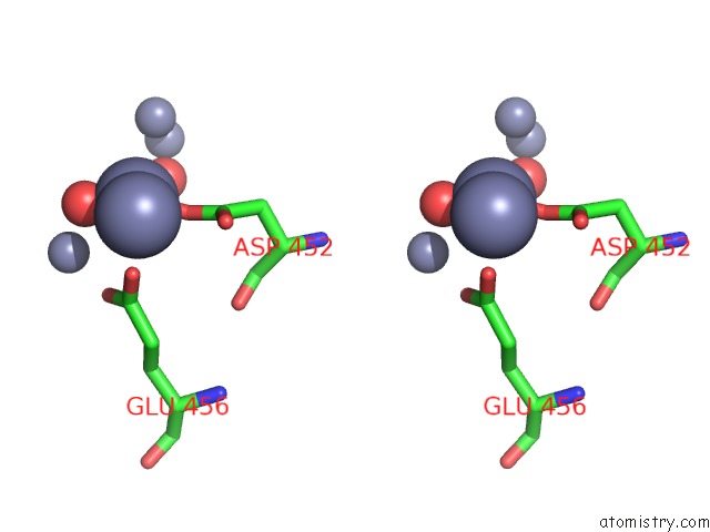
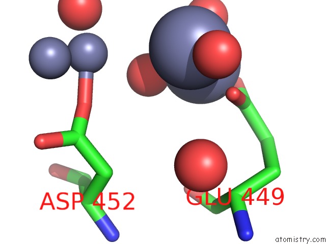
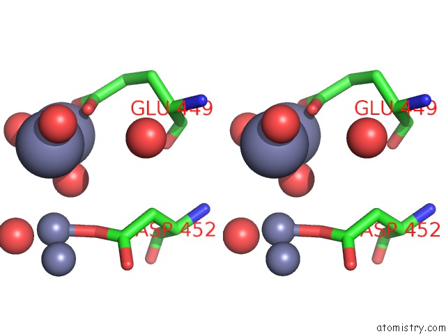
Zinc binding site 2 out of 7 in 2akf
Go back to
Zinc binding site 2 out
of 7 in the Crystal Structure of the Coiled-Coil Domain of Coronin 1
Mono view
Stereo pair view
Mono view
Stereo pair view
A full contact list of Zinc with other atoms in the Zn binding
site number 2 of Crystal Structure of the Coiled-Coil Domain of Coronin 1 within 5.0Å range:
|
Zinc binding site 3 out of 7 in 2akf
Go back to
Zinc binding site 3 out
of 7 in the Crystal Structure of the Coiled-Coil Domain of Coronin 1
Mono view
Stereo pair view
Mono view
Stereo pair view
A full contact list of Zinc with other atoms in the Zn binding
site number 3 of Crystal Structure of the Coiled-Coil Domain of Coronin 1 within 5.0Å range:
|
Zinc binding site 4 out of 7 in 2akf
Go back to
Zinc binding site 4 out
of 7 in the Crystal Structure of the Coiled-Coil Domain of Coronin 1
Mono view
Stereo pair view
Mono view
Stereo pair view
A full contact list of Zinc with other atoms in the Zn binding
site number 4 of Crystal Structure of the Coiled-Coil Domain of Coronin 1 within 5.0Å range:
|
Zinc binding site 5 out of 7 in 2akf
Go back to
Zinc binding site 5 out
of 7 in the Crystal Structure of the Coiled-Coil Domain of Coronin 1
Mono view
Stereo pair view
Mono view
Stereo pair view
A full contact list of Zinc with other atoms in the Zn binding
site number 5 of Crystal Structure of the Coiled-Coil Domain of Coronin 1 within 5.0Å range:
|
Zinc binding site 6 out of 7 in 2akf
Go back to
Zinc binding site 6 out
of 7 in the Crystal Structure of the Coiled-Coil Domain of Coronin 1
Mono view
Stereo pair view
Mono view
Stereo pair view
A full contact list of Zinc with other atoms in the Zn binding
site number 6 of Crystal Structure of the Coiled-Coil Domain of Coronin 1 within 5.0Å range:
|
Zinc binding site 7 out of 7 in 2akf
Go back to
Zinc binding site 7 out
of 7 in the Crystal Structure of the Coiled-Coil Domain of Coronin 1
Mono view
Stereo pair view
Mono view
Stereo pair view
A full contact list of Zinc with other atoms in the Zn binding
site number 7 of Crystal Structure of the Coiled-Coil Domain of Coronin 1 within 5.0Å range:
|
Reference:
R.A.Kammerer,
D.Kostrewa,
P.Progias,
S.Honnappa,
D.Avila,
A.Lustig,
F.K.Winkler,
J.Pieters,
M.O.Steinmetz.
A Conserved Trimerization Motif Controls the Topology of Short Coiled Coils Proc.Natl.Acad.Sci.Usa V. 102 13891 2005.
ISSN: ISSN 0027-8424
PubMed: 16172398
DOI: 10.1073/PNAS.0502390102
Page generated: Wed Oct 16 21:40:22 2024
ISSN: ISSN 0027-8424
PubMed: 16172398
DOI: 10.1073/PNAS.0502390102
Last articles
Zn in 9MJ5Zn in 9HNW
Zn in 9G0L
Zn in 9FNE
Zn in 9DZN
Zn in 9E0I
Zn in 9D32
Zn in 9DAK
Zn in 8ZXC
Zn in 8ZUF