Zinc »
PDB 2afo-2aqr »
2afo »
Zinc in PDB 2afo: Crystal Structure of Human Glutaminyl Cyclase at pH 8.0
Enzymatic activity of Crystal Structure of Human Glutaminyl Cyclase at pH 8.0
All present enzymatic activity of Crystal Structure of Human Glutaminyl Cyclase at pH 8.0:
2.3.2.5;
2.3.2.5;
Protein crystallography data
The structure of Crystal Structure of Human Glutaminyl Cyclase at pH 8.0, PDB code: 2afo
was solved by
K.F.Huang,
Y.L.Liu,
W.J.Cheng,
T.P.Ko,
A.H.J.Wang,
with X-Ray Crystallography technique. A brief refinement statistics is given in the table below:
| Resolution Low / High (Å) | 30.00 / 2.35 |
| Space group | H 3 2 |
| Cell size a, b, c (Å), α, β, γ (°) | 118.988, 118.988, 332.258, 90.00, 90.00, 120.00 |
| R / Rfree (%) | 18.5 / 21.6 |
Zinc Binding Sites:
The binding sites of Zinc atom in the Crystal Structure of Human Glutaminyl Cyclase at pH 8.0
(pdb code 2afo). This binding sites where shown within
5.0 Angstroms radius around Zinc atom.
In total 2 binding sites of Zinc where determined in the Crystal Structure of Human Glutaminyl Cyclase at pH 8.0, PDB code: 2afo:
Jump to Zinc binding site number: 1; 2;
In total 2 binding sites of Zinc where determined in the Crystal Structure of Human Glutaminyl Cyclase at pH 8.0, PDB code: 2afo:
Jump to Zinc binding site number: 1; 2;
Zinc binding site 1 out of 2 in 2afo
Go back to
Zinc binding site 1 out
of 2 in the Crystal Structure of Human Glutaminyl Cyclase at pH 8.0
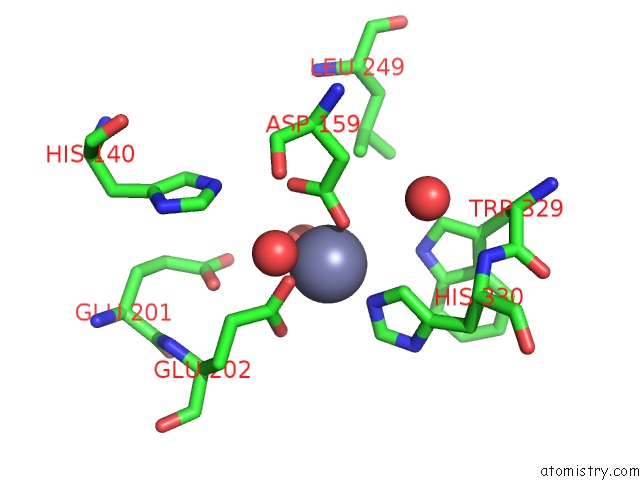
Mono view
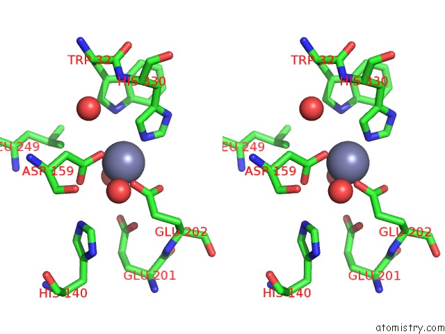
Stereo pair view
Mono view
Stereo pair view
A full contact list of Zinc with other atoms in the Zn binding
site number 1 of Crystal Structure of Human Glutaminyl Cyclase at pH 8.0 within 5.0Å range:
|
Zinc binding site 2 out of 2 in 2afo
Go back to
Zinc binding site 2 out
of 2 in the Crystal Structure of Human Glutaminyl Cyclase at pH 8.0
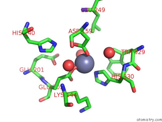
Mono view
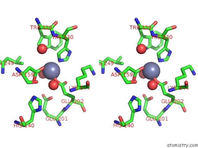
Stereo pair view
Mono view
Stereo pair view
A full contact list of Zinc with other atoms in the Zn binding
site number 2 of Crystal Structure of Human Glutaminyl Cyclase at pH 8.0 within 5.0Å range:
|
Reference:
K.F.Huang,
Y.L.Liu,
W.J.Cheng,
T.P.Ko,
A.H.Wang.
Crystal Structures of Human Glutaminyl Cyclase, An Enzyme Responsible For Protein N-Terminal Pyroglutamate Formation Proc.Natl.Acad.Sci.Usa V. 102 13117 2005.
ISSN: ISSN 0027-8424
PubMed: 16135565
DOI: 10.1073/PNAS.0504184102
Page generated: Wed Oct 16 21:37:25 2024
ISSN: ISSN 0027-8424
PubMed: 16135565
DOI: 10.1073/PNAS.0504184102
Last articles
Zn in 9MJ5Zn in 9HNW
Zn in 9G0L
Zn in 9FNE
Zn in 9DZN
Zn in 9E0I
Zn in 9D32
Zn in 9DAK
Zn in 8ZXC
Zn in 8ZUF