Zinc »
PDB 2a2q-2afm »
2a8a »
Zinc in PDB 2a8a: Crystal Structure of Clostridium Botulinum Neurotoxin Serotype F Light Chain
Enzymatic activity of Crystal Structure of Clostridium Botulinum Neurotoxin Serotype F Light Chain
All present enzymatic activity of Crystal Structure of Clostridium Botulinum Neurotoxin Serotype F Light Chain:
3.4.24.69;
3.4.24.69;
Protein crystallography data
The structure of Crystal Structure of Clostridium Botulinum Neurotoxin Serotype F Light Chain, PDB code: 2a8a
was solved by
R.Agarwal,
T.Binz,
S.Swaminathan,
with X-Ray Crystallography technique. A brief refinement statistics is given in the table below:
| Resolution Low / High (Å) | 47.67 / 2.00 |
| Space group | C 1 2 1 |
| Cell size a, b, c (Å), α, β, γ (°) | 63.413, 79.463, 89.660, 90.00, 110.01, 90.00 |
| R / Rfree (%) | 22.6 / 26.5 |
Other elements in 2a8a:
The structure of Crystal Structure of Clostridium Botulinum Neurotoxin Serotype F Light Chain also contains other interesting chemical elements:
| Cadmium | (Cd) | 2 atoms |
Zinc Binding Sites:
The binding sites of Zinc atom in the Crystal Structure of Clostridium Botulinum Neurotoxin Serotype F Light Chain
(pdb code 2a8a). This binding sites where shown within
5.0 Angstroms radius around Zinc atom.
In total only one binding site of Zinc was determined in the Crystal Structure of Clostridium Botulinum Neurotoxin Serotype F Light Chain, PDB code: 2a8a:
In total only one binding site of Zinc was determined in the Crystal Structure of Clostridium Botulinum Neurotoxin Serotype F Light Chain, PDB code: 2a8a:
Zinc binding site 1 out of 1 in 2a8a
Go back to
Zinc binding site 1 out
of 1 in the Crystal Structure of Clostridium Botulinum Neurotoxin Serotype F Light Chain
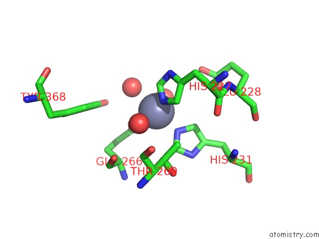
Mono view
Stereo pair view
Mono view
Stereo pair view
A full contact list of Zinc with other atoms in the Zn binding
site number 1 of Crystal Structure of Clostridium Botulinum Neurotoxin Serotype F Light Chain within 5.0Å range:
|
Reference:
R.Agarwal,
T.Binz,
S.Swaminathan.
Structural Analysis of Botulinum Neurotoxin Serotype F Light Chain: Implications on Substrate Binding and Inhibitor Design Biochemistry V. 44 11758 2005.
ISSN: ISSN 0006-2960
PubMed: 16128577
DOI: 10.1021/BI0510072
Page generated: Wed Oct 16 21:33:17 2024
ISSN: ISSN 0006-2960
PubMed: 16128577
DOI: 10.1021/BI0510072
Last articles
Zn in 9MJ5Zn in 9HNW
Zn in 9G0L
Zn in 9FNE
Zn in 9DZN
Zn in 9E0I
Zn in 9D32
Zn in 9DAK
Zn in 8ZXC
Zn in 8ZUF