Zinc »
PDB 2a2q-2afm »
2a3g »
Zinc in PDB 2a3g: The Structure of T6 Bovine Insulin
Protein crystallography data
The structure of The Structure of T6 Bovine Insulin, PDB code: 2a3g
was solved by
G.D.Smith,
W.A.Pangborn,
R.H.Blessing,
with X-Ray Crystallography technique. A brief refinement statistics is given in the table below:
| Resolution Low / High (Å) | 30.50 / 2.25 |
| Space group | H 3 |
| Cell size a, b, c (Å), α, β, γ (°) | 82.554, 82.554, 33.756, 90.00, 90.00, 120.00 |
| R / Rfree (%) | 16.2 / 21.9 |
Zinc Binding Sites:
The binding sites of Zinc atom in the The Structure of T6 Bovine Insulin
(pdb code 2a3g). This binding sites where shown within
5.0 Angstroms radius around Zinc atom.
In total 2 binding sites of Zinc where determined in the The Structure of T6 Bovine Insulin, PDB code: 2a3g:
Jump to Zinc binding site number: 1; 2;
In total 2 binding sites of Zinc where determined in the The Structure of T6 Bovine Insulin, PDB code: 2a3g:
Jump to Zinc binding site number: 1; 2;
Zinc binding site 1 out of 2 in 2a3g
Go back to
Zinc binding site 1 out
of 2 in the The Structure of T6 Bovine Insulin
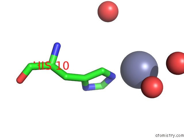
Mono view
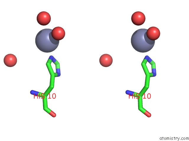
Stereo pair view
Mono view
Stereo pair view
A full contact list of Zinc with other atoms in the Zn binding
site number 1 of The Structure of T6 Bovine Insulin within 5.0Å range:
|
Zinc binding site 2 out of 2 in 2a3g
Go back to
Zinc binding site 2 out
of 2 in the The Structure of T6 Bovine Insulin
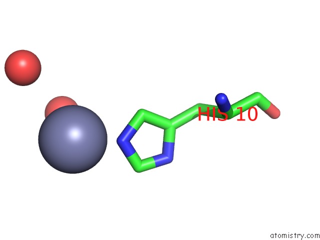
Mono view
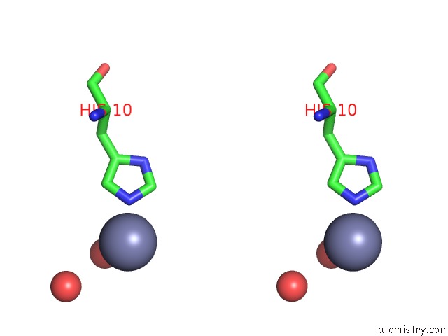
Stereo pair view
Mono view
Stereo pair view
A full contact list of Zinc with other atoms in the Zn binding
site number 2 of The Structure of T6 Bovine Insulin within 5.0Å range:
|
Reference:
G.D.Smith,
W.A.Pangborn,
R.H.Blessing.
The Structure of T6 Bovine Insulin. Acta Crystallogr.,Sect.D V. 61 1476 2005.
ISSN: ISSN 0907-4449
PubMed: 16239724
DOI: 10.1107/S0907444905025771
Page generated: Wed Oct 16 21:29:22 2024
ISSN: ISSN 0907-4449
PubMed: 16239724
DOI: 10.1107/S0907444905025771
Last articles
Zn in 9MJ5Zn in 9HNW
Zn in 9G0L
Zn in 9FNE
Zn in 9DZN
Zn in 9E0I
Zn in 9D32
Zn in 9DAK
Zn in 8ZXC
Zn in 8ZUF