Zinc »
PDB 1z5r-1zfo »
1z9p »
Zinc in PDB 1z9p: X-Ray Structure of A Cu-Zn Superoxide Dismutase From Haemophilus Ducreyi
Enzymatic activity of X-Ray Structure of A Cu-Zn Superoxide Dismutase From Haemophilus Ducreyi
All present enzymatic activity of X-Ray Structure of A Cu-Zn Superoxide Dismutase From Haemophilus Ducreyi:
1.15.1.1;
1.15.1.1;
Protein crystallography data
The structure of X-Ray Structure of A Cu-Zn Superoxide Dismutase From Haemophilus Ducreyi, PDB code: 1z9p
was solved by
K.Djinovic Carugo,
I.Toeroe,
with X-Ray Crystallography technique. A brief refinement statistics is given in the table below:
| Resolution Low / High (Å) | 19.32 / 1.50 |
| Space group | C 1 2 1 |
| Cell size a, b, c (Å), α, β, γ (°) | 71.410, 63.990, 73.970, 90.00, 118.04, 90.00 |
| R / Rfree (%) | 17 / 21.2 |
Other elements in 1z9p:
The structure of X-Ray Structure of A Cu-Zn Superoxide Dismutase From Haemophilus Ducreyi also contains other interesting chemical elements:
| Copper | (Cu) | 2 atoms |
Zinc Binding Sites:
The binding sites of Zinc atom in the X-Ray Structure of A Cu-Zn Superoxide Dismutase From Haemophilus Ducreyi
(pdb code 1z9p). This binding sites where shown within
5.0 Angstroms radius around Zinc atom.
In total 2 binding sites of Zinc where determined in the X-Ray Structure of A Cu-Zn Superoxide Dismutase From Haemophilus Ducreyi, PDB code: 1z9p:
Jump to Zinc binding site number: 1; 2;
In total 2 binding sites of Zinc where determined in the X-Ray Structure of A Cu-Zn Superoxide Dismutase From Haemophilus Ducreyi, PDB code: 1z9p:
Jump to Zinc binding site number: 1; 2;
Zinc binding site 1 out of 2 in 1z9p
Go back to
Zinc binding site 1 out
of 2 in the X-Ray Structure of A Cu-Zn Superoxide Dismutase From Haemophilus Ducreyi
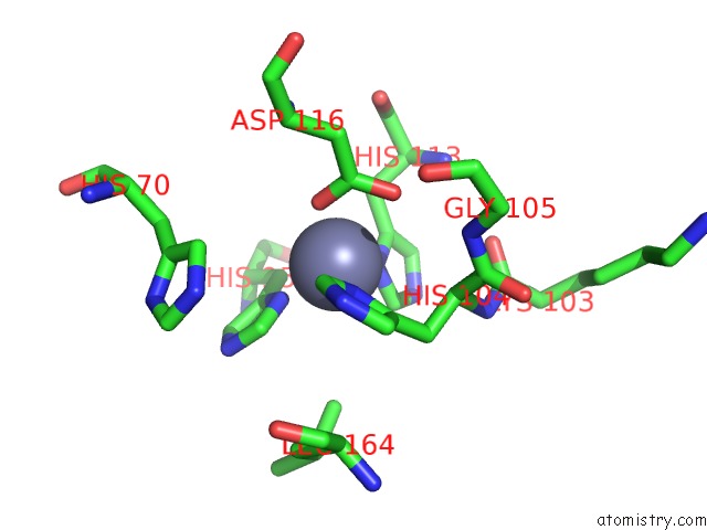
Mono view
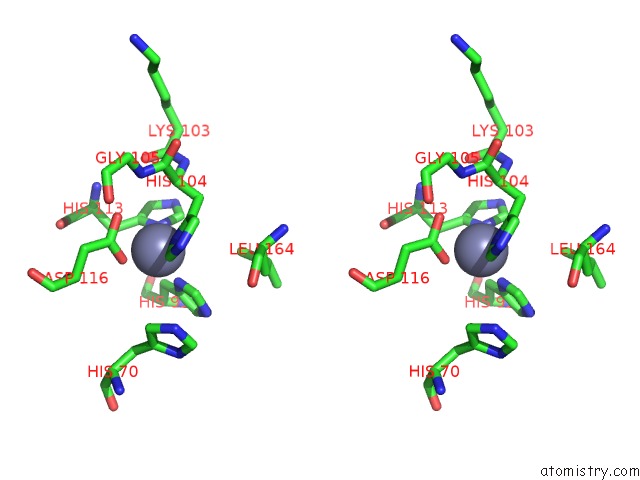
Stereo pair view
Mono view
Stereo pair view
A full contact list of Zinc with other atoms in the Zn binding
site number 1 of X-Ray Structure of A Cu-Zn Superoxide Dismutase From Haemophilus Ducreyi within 5.0Å range:
|
Zinc binding site 2 out of 2 in 1z9p
Go back to
Zinc binding site 2 out
of 2 in the X-Ray Structure of A Cu-Zn Superoxide Dismutase From Haemophilus Ducreyi
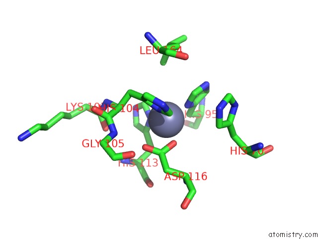
Mono view
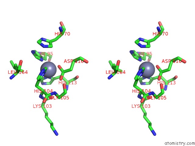
Stereo pair view
Mono view
Stereo pair view
A full contact list of Zinc with other atoms in the Zn binding
site number 2 of X-Ray Structure of A Cu-Zn Superoxide Dismutase From Haemophilus Ducreyi within 5.0Å range:
|
Reference:
I.Toro,
C.Petrutz,
F.Pacello,
M.D'orazio,
A.Battistoni,
K.Djinovic-Carugo.
Structural Basis of Heme Binding in the Cu,Zn Superoxide Dismutase From Haemophilus Ducreyi. J.Mol.Biol. V. 386 406 2009.
ISSN: ISSN 0022-2836
PubMed: 19103206
DOI: 10.1016/J.JMB.2008.12.004
Page generated: Wed Aug 20 00:49:58 2025
ISSN: ISSN 0022-2836
PubMed: 19103206
DOI: 10.1016/J.JMB.2008.12.004
Last articles
Zn in 2MLRZn in 2MLS
Zn in 2MMI
Zn in 2MMH
Zn in 2MKE
Zn in 2MIU
Zn in 2MKD
Zn in 2MKN
Zn in 2MJC
Zn in 2MIQ