Zinc »
PDB 1z5r-1zfo »
1z7h »
Zinc in PDB 1z7h: 2.3 Angstrom Crystal Structure of Tetanus Neurotoxin Light Chain
Enzymatic activity of 2.3 Angstrom Crystal Structure of Tetanus Neurotoxin Light Chain
All present enzymatic activity of 2.3 Angstrom Crystal Structure of Tetanus Neurotoxin Light Chain:
3.4.24.68;
3.4.24.68;
Protein crystallography data
The structure of 2.3 Angstrom Crystal Structure of Tetanus Neurotoxin Light Chain, PDB code: 1z7h
was solved by
M.A.Breidenbach,
A.T.Brunger,
with X-Ray Crystallography technique. A brief refinement statistics is given in the table below:
| Resolution Low / High (Å) | 38.76 / 2.30 |
| Space group | C 2 2 2 |
| Cell size a, b, c (Å), α, β, γ (°) | 105.350, 176.860, 57.230, 90.00, 90.00, 90.00 |
| R / Rfree (%) | 21.2 / 21.6 |
Zinc Binding Sites:
The binding sites of Zinc atom in the 2.3 Angstrom Crystal Structure of Tetanus Neurotoxin Light Chain
(pdb code 1z7h). This binding sites where shown within
5.0 Angstroms radius around Zinc atom.
In total 2 binding sites of Zinc where determined in the 2.3 Angstrom Crystal Structure of Tetanus Neurotoxin Light Chain, PDB code: 1z7h:
Jump to Zinc binding site number: 1; 2;
In total 2 binding sites of Zinc where determined in the 2.3 Angstrom Crystal Structure of Tetanus Neurotoxin Light Chain, PDB code: 1z7h:
Jump to Zinc binding site number: 1; 2;
Zinc binding site 1 out of 2 in 1z7h
Go back to
Zinc binding site 1 out
of 2 in the 2.3 Angstrom Crystal Structure of Tetanus Neurotoxin Light Chain
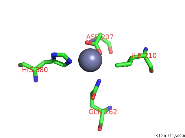
Mono view
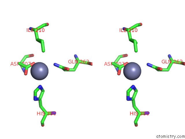
Stereo pair view
Mono view
Stereo pair view
A full contact list of Zinc with other atoms in the Zn binding
site number 1 of 2.3 Angstrom Crystal Structure of Tetanus Neurotoxin Light Chain within 5.0Å range:
|
Zinc binding site 2 out of 2 in 1z7h
Go back to
Zinc binding site 2 out
of 2 in the 2.3 Angstrom Crystal Structure of Tetanus Neurotoxin Light Chain
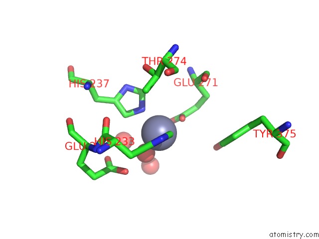
Mono view
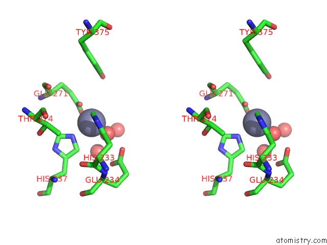
Stereo pair view
Mono view
Stereo pair view
A full contact list of Zinc with other atoms in the Zn binding
site number 2 of 2.3 Angstrom Crystal Structure of Tetanus Neurotoxin Light Chain within 5.0Å range:
|
Reference:
M.A.Breidenbach,
A.T.Brunger.
2.3A Crystal Structure of Tetanus Neurotoxin Light Chain Biochemistry V. 44 7450 2005.
ISSN: ISSN 0006-2960
PubMed: 15895988
DOI: 10.1021/BI050262J
Page generated: Wed Aug 20 00:48:28 2025
ISSN: ISSN 0006-2960
PubMed: 15895988
DOI: 10.1021/BI050262J
Last articles
Zn in 2N94Zn in 2N8R
Zn in 2N6J
Zn in 2N26
Zn in 2N5K
Zn in 2N1A
Zn in 2N25
Zn in 2N1U
Zn in 2MZZ
Zn in 2MZI